. 2018 Oct 25;16(1):295.
doi: 10.1186/s12967-018-1670-9.

An increase in myocardial 18-fluorodeoxyglucose uptake is associated with left ventricular ejection fraction decline in Hodgkin lymphoma patients treated with anthracycline

Affiliations

An increase in myocardial 18-fluorodeoxyglucose uptake is associated with left ventricular ejection fraction decline in Hodgkin lymphoma patients treated with anthracycline

Matteo Sarocchi et al. J Transl Med. .

Abstract

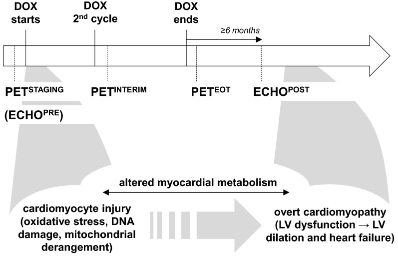
Background: Doxorubicin (DOX)-based chemotherapy for Hodgkin lymphoma (HL) yields excellent disease-free survival, but poses a substantial risk of subsequent left ventricular (LV) dysfunction and heart failure, typically with delayed onset. At the cellular level, this cardiotoxicity includes deranged cardiac glucose metabolism.

Methods: By reviewing the hospital records from January 2008 through December 2016, we selected HL patients meeting the following criteria: ≥ 18 year-old; first-line DOX-containing chemotherapy; no diabetes and apparent cardiovascular disease; 18-fluoro-deoxyglucose positron emission tomography (18FDG-PET) scans before treatment (PETSTAGING), after 2 cycles (PETINTERIM) and at the end of treatment (PETEOT); at least one echocardiography ≥ 6 months after chemotherapy completion (ECHOPOST). We then evaluated the changes in LV 18FDG standardized uptake values (SUV) during the course of DOX therapy, and the relationship between LV-SUV and LV ejection fraction (LVEF), as calculated from the LV diameters in the echocardiography reports with the Teicholz formula.

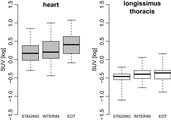
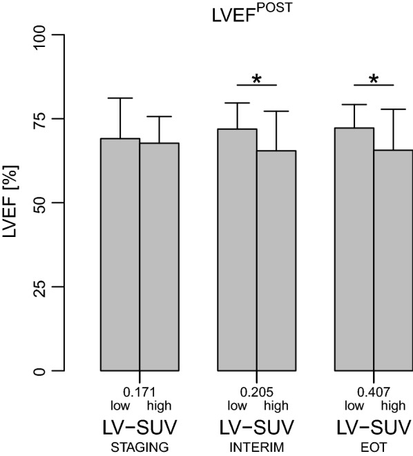
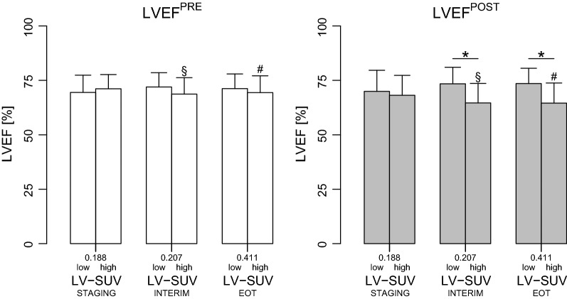
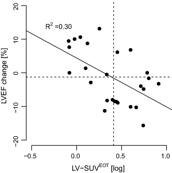
Results: Forty-three patients (35 ± 13 year-old, 58% males) were included in the study, with 26 (60%) also having a baseline echocardiography available (ECHOPRE). LV-SUV gradually increased from PETSTAGING (log-transformed mean 0.20 ± 0.27) to PETINTERIM (0.27 ± 0.35) to PETEOT (0.30 ± 0.41; P for trend < 0.001). ECHOPOST was performed 22 ± 17 months after DOX chemotherapy. Mean LVEF was normal (68.8 ± 10.3%) and only three subjects (7%) faced a drop below the upper normal limit of 53%. However, when patients were categorized by median LV-SUV, LVEF at ECHOPOST resulted significantly lower in those with LV-SUV above than below the median value at both PETINTERIM (65.5 ± 11.8% vs. 71.9 ± 7.8%, P = 0.04) and PETEOT (65.6 ± 12.2% vs. 72.2 ± 7.0%, P = 0.04). This was also the case when only patients with ECHOPRE and ECHOPOST were considered (LVEF at ECHOPOST 64.7 ± 8.9% vs. 73.4 ± 7.6%, P = 0.01 and 64.6 ± 9.3% vs. 73.5 ± 7.0%, P = 0.01 for those with LV-SUV above vs. below the median at PETINTERIM and PETEOT, respectively). Furthermore, the difference between LVEF at ECHOPRE and ECHOPOST was inversely correlated with LV-SUV at PETEOT (P < 0.01, R2 = - 0.30).

Conclusions: DOX-containing chemotherapy causes an increase in cardiac 18FDG uptake, which is associated with a decline in LVEF. Future studies are warranted to understand the molecular basis and the potential clinical implications of this observation.

Keywords: 18FDG-PET; Cardiotoxicity; Doxorubicin; Heart failure; Left ventricular dysfunction.


Figures

Fig. 1
Fig. 1
Study design and stages of anthracycline cardiotoxicity. A schematic of the timing of the 18FDG positron emission tomography (18FDG-PET) scans and echocardiograms taken into analysis is depicted in the upper panel, while the corresponding stages of anthracycline-related cardiotoxicity are presented in the lower part. DOX: doxorubicin-containing chemotherapy; ECHOPRE: echocardiography at baseline (available only in a subgroup of patients); ECHOPOST: echocardiography after completion of DOX chemotherapy; PETSTAGING: 18FDG-PET before treatment; PETINTERIM: 18FDG-PET after 2 cycles of doxorubicin; PETEOT: 18FDG-PET at the end of treatment; LV: left ventricular
Fig. 2
Fig. 2
18FDG uptake during doxorubicin treatment in the myocardium and in the longissimus thoracis muscle. Boxes are median and interquartile ranges of left ventricular and skeletal muscle 18FDG standardized uptake values (SUV) at the indicated 18FDG positron emission tomography (18FDG-PET) scans. Vertical bars indicate the highest and lowest SUV at each time point. Repeated measures ANOVA P for trend = 0.0007 and 0.02 for SUV in the heart and longissimus thoracis, respectively. STAGING: 18FDG-PET before treatment; INTERIM: 18FDG-PET after 2 cycles of doxorubicin; EOT: 18FDG-PET at the end of treatment
Fig. 3
Fig. 3
Left ventricular ejection fraction according to categories of 18FDG uptake in patients with echocardiography at follow-up. Left ventricular ejection fraction (LVEF), as assessed by echocardiography performed after completion of anthracycline chemotherapy, in patients with myocardial 18FDG standardized uptake values (LV-SUV) below or above the median value (low and high, respectively) measured at each 18FDG positron emission tomography (18FDG-PET) scan. For each PET time point, LVEF was compared between patients with LV-SUV below vs. above the median value by unpaired t-test. * indicates P < 0.05. LV-SUVSTAGING: LV-SUV at the 18FDG-PET scan before treatment; LV-SUVINTERIM: LV-SUV at the 18FDG-PET performed after 2 cycles of doxorubicin; LV-SUVEOT: LV-SUV at the 18FDG-PET performed at the end of chemotherapy
Fig. 4
Fig. 4
Left ventricular ejection fraction according to categories of 18FDG uptake in patients with echocardiography at both baseline and follow-up. Left ventricular ejection fraction (LVEF) at the baseline and follow-up echocardiography (LVEFPRE and LVEFPOST, respectively) in patients with myocardial 18FDG standardized uptake values (LV-SUV) below or above the median value (low and high, respectively) at each 18FDG positron emission tomography (18FDG-PET) scan. LVEF was compared between patients with LV-SUV below vs. above the median value at each PET time point by unpaired t-test and * is P < 0.05. LVEFPRE and LVEFPOST for each subgroup of patients with LV-SUV below or above the median value (e.g. LVEFPRE and LVEFPOST in subjects with high LV-SUV at EOT) were compared by paired t-test; § and # indicate P < 0.05 and < 0.01, respectively. LV-SUVSTAGING: LV-SUV at the 18FDG-PET scan before treatment; LV-SUVINTERIM: LV-SUV at the 18FDG-PET performed after 2 cycles of doxorubicin; LV-SUVEOT: LV-SUV at the 18FDG-PET performed at the end of chemotherapy
Fig. 5
Fig. 5
Relationship between left ventricular ejection fraction change and 18FDG uptake in the subsets of patients with baseline and follow-up echocardiography data available. LVEF: left ventricular ejection fraction; LV-SUVEOT: LV 18FDG standardized uptake value at positron emission tomography performed at the end of anthracycline chemotherapy

References

    1. Ansell SM. Hodgkin lymphoma: 2014 update on diagnosis, risk-stratification, and management. Am J Hematol. 2014;89(7):771–779. doi: 10.1002/ajh.23750. - DOI - PubMed
    1. Barrington SF, Kirkwood AA, Franceschetto A, et al. PET-CT for staging and early response: results from the response-adapted therapy in advanced Hodgkin lymphoma study. Blood. 2016;127(12):1531–1538. doi: 10.1182/blood-2015-11-679407. - DOI - PubMed
    1. Smith LA, Cornelius VR, Plummer CJ, et al. Cardiotoxicity of anthracycline agents for the treatment of cancer: systematic review and meta-analysis of randomised controlled trials. BMC Cancer. 2010;10(1):337. doi: 10.1186/1471-2407-10-337. - DOI - PMC - PubMed
    1. Bhakta N, Liu Q, Yeo F, et al. Cumulative burden of cardiovascular morbidity in paediatric, adolescent, and young adult survivors of Hodgkin’s lymphoma: an analysis from the St Jude Lifetime Cohort Study. Lancet Oncol. 2016;17(9):1325–1334. doi: 10.1016/S1470-2045(16)30215-7. - DOI - PMC - PubMed
    1. van Nimwegen FA, Schaapveld M, Janus CPM, et al. Cardiovascular disease after Hodgkin lymphoma treatment: 40-year disease risk. JAMA Intern Med. 2015;175(6):1007–1017. doi: 10.1001/jamainternmed.2015.1180. - DOI - PubMed

Publication types