. 2018 Oct 25;13(10):e0206161.
doi: 10.1371/journal.pone.0206161. eCollection 2018.

Dissociation of Rpb4 from RNA polymerase II is important for yeast functionality

Affiliations

Dissociation of Rpb4 from RNA polymerase II is important for yeast functionality

Lea Duek et al. PLoS One. .

Abstract

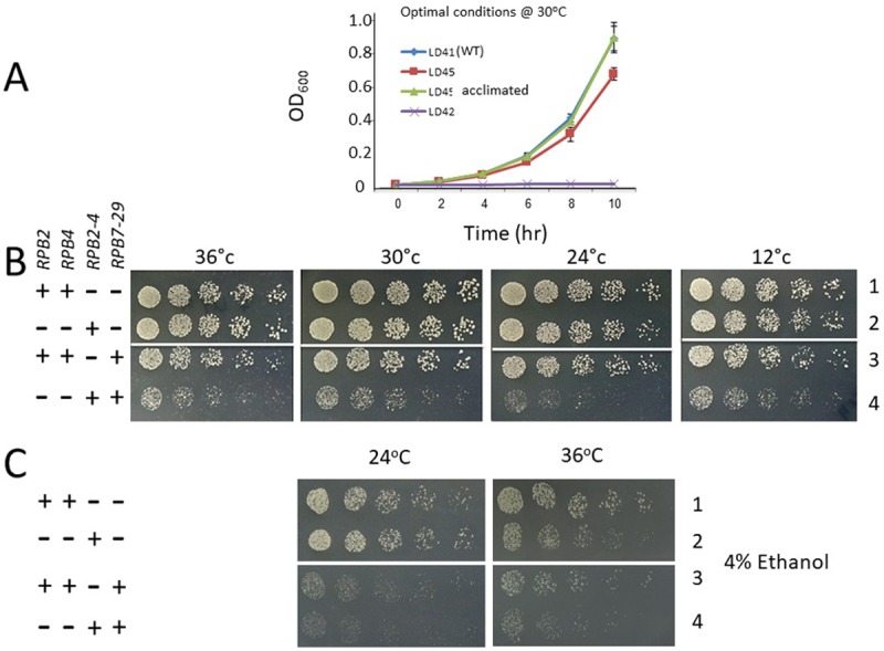
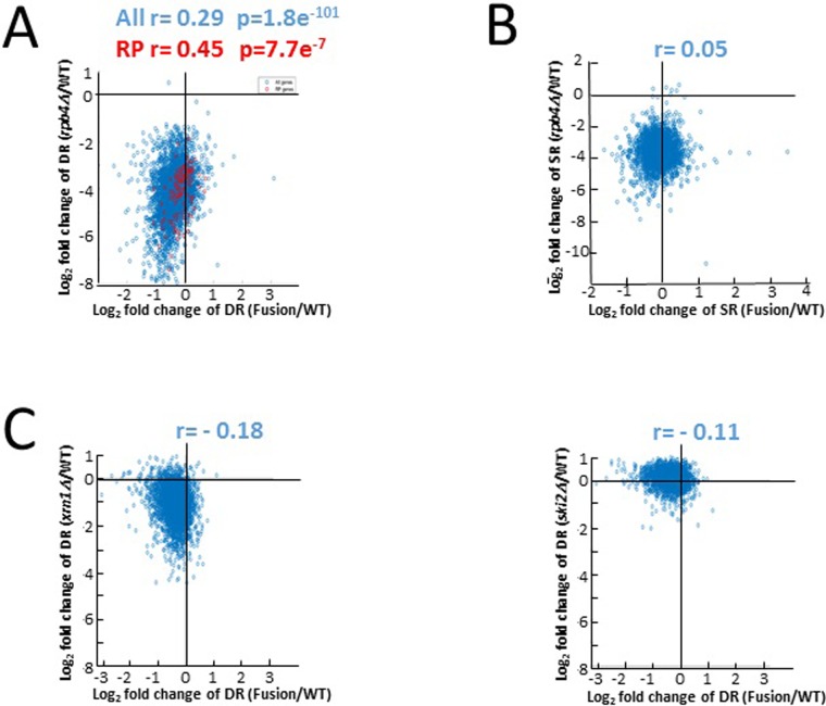
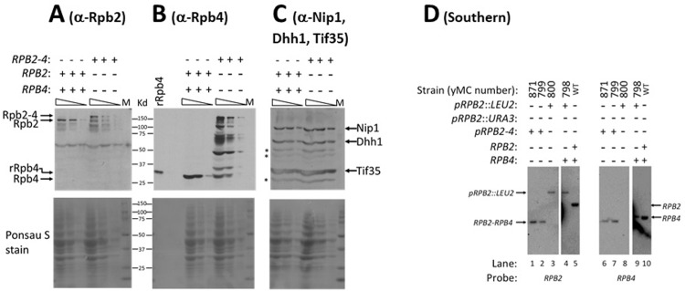
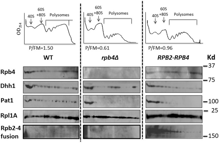
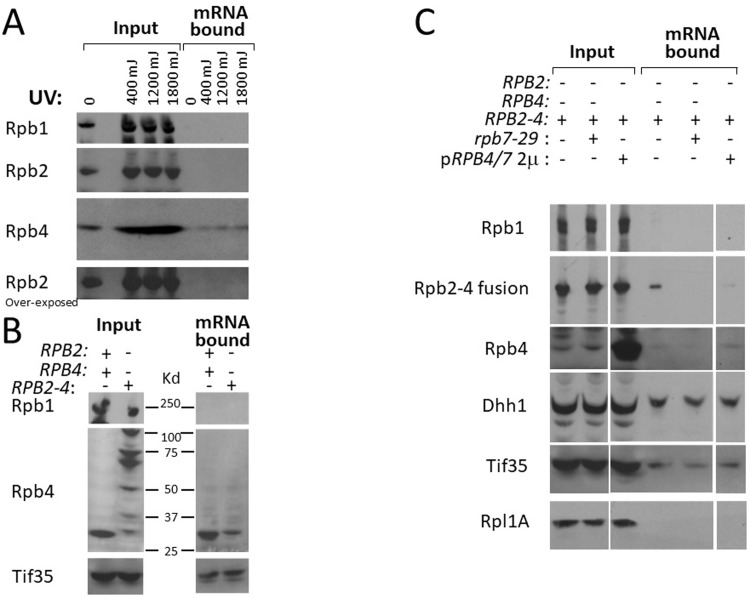
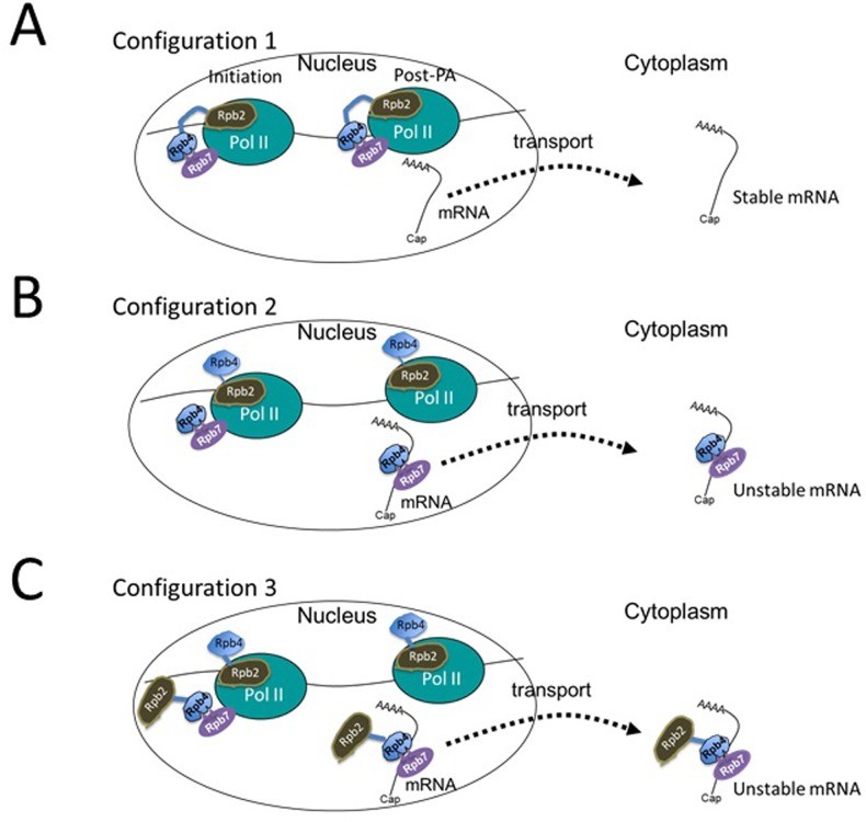
Rpb4 is an RNA polymerase II (Pol II) subunit that binds Pol II transcripts co-transcriptionally, accompanies them to the cytoplasm and modulates mRNA export, translation and decay by interacting with cytoplasmic RNA modulators. The importance of the cytoplasmic roles of Rpb4 was challenged by a study reporting that the phenotype of rpb2Δ rpb4Δ cells can be rescued by an Rpb2-Rpb4 fusion protein, assuming that its Rpb4 moiety cannot dissociate from Pol II and functions in the cytoplasm. Here we demonstrate that although the fusion protein supports normal transcription, it adversely affects mRNA decay, cell proliferation and adaptability-e.g., response to stress. These defects are similar, albeit milder, than the defects that characterize rpb4Δ cells. At least two mechanisms alleviate the deleterious effect of the fusion protein. First, a portion of this fusion protein is cleaved into free Rpb2 and Rpb4. The free Rpb4 is functional, as it binds mRNAs and polysomes, like WT Rpb4. Second, the fusion protein is also capable of binding poly(A)+ mRNAs in the cytoplasm, in an Rpb7-mediated manner, probably complementing the functions of the diminished Rpb4. Collectively, normal coupling between mRNA synthesis and decay requires wild-type configuration of Rpb4, and fusing Rpb4 to Rpb2 compromises this coupling.


Conflict of interest statement

The authors have declared that no competing interests exist.

Figures

Fig 1
Fig 1. Expression of the RPB2-RPB4 fusion gene compromises cell proliferation.
(A) The yLD45 strain was grown in two parallel cultures in SC medium lacking leucine. One culture was supplemented with 10 μg/ml doxycycline (named "yLD45 acclimated") to repress expression of the Tet-off RPB2. After ~60 generations of growth in logarithmic phase (with intermittent dilutions), the two cultures were diluted into fresh medium containing doxycycline for 24 h (relatively short) acclimation to the drug. Cultures were then diluted, in triplicate, into fresh medium containing doxycycline and proliferation rate was measured. Data are presented as means ± SD (n = 3). yLD42 contains Tet-off-RPB2 as the sole source of Rpb2, but otherwise is WT; yLD41 is a derivative of yLD42 that expresses pRPB2::LEU2 and therefore proliferates as WT even in the presence of doxycycline. (B) Expression of RPB2-RPB4 is deleterious in cells that overexpress rpb7-29. Spot test of 5-fold serial dilution of yMC798, yMC871, yMC923 and yMC924 (from top to bottom) grown on selective plates at the indicated temperatures. The first spot of each strain was of 5000 cells. (C) Spot test of 5-fold serial dilution of the indicated strains (same as in B) grown on selective plates containing 4% ethanol as the main carbon source. The first spot of each strain was of 5000 cells. The growth temperature is indicated above each panel.
Fig 2
Fig 2. Correlation between mRNA synthesis and decay rates in RPB2-RPB4 cells against those in rpb4Δ cells.
mRNA synthesis rates (SRs) and decay rates (DRs) data of rpb4Δ and of RPB2-RPB4 strains were taken from Schulz et al. [15]; DR data of the other strains were taken from [39]. (A) Fold changes in DRs for rpb4Δ cells (log2 folds of rpb4Δ/WT, y axis) against fold changes in DRs for RPB2-RPB4 cells (log2 folds of RPB2-RPB4/WT, x axis). Each spot corresponds to one mRNA. Red spot represent mRNAs encoding ribosomal proteins whose decay rates are more dependent on Rpb4 than other mRNAs [21]. Spearman correlation was used to determine the correlation. (B) Correlation of SRs. Analysis similar to that in A was performed on SRs data. (C) Correlation analyses of DRs for xrn1Δ and ski2Δ strains against RPB2-RPB4 strain were performed as in A. All strains are isogenic. See also S2 Fig.
Fig 3
Fig 3. RPB2-RPB4 cells do not contain other source of Rpb2 and Rpb4 and express Rpb2-Rbp4 fusion protein as well as Rpb2 and Rpb4 proteins.
(A-C) Cell lysates of the indicated strains were loaded on polyacrylamide gel; each lysate was loaded in 3 lanes, containing either 200 μg, 70 μg or 20 μl of protein/lane. Three replicates, each containing 6 lanes, were thus loaded and analyzed by Western blotting assay; each membrane was reacted with either anti-Rpb2 Abs (A) or anti-Rpb4 Abs (B), or with a combination of anti-Nip1 + anti-Dhh1 + anti-Tif35 (C). Thirty ng of recombinant Rpb4-HISx6 (rRpb4), expressed in E. coli, was loaded in panel A to mark the position of Rpb4 (note that the rRpb4 migrated slower than Rpb4 due to an HISx6 tag that was used for its purification). Ponsau S stain of the membranes is shown underneath each respective panel. The pre-stained size marker (BioRad) is also shown. Asterisk indicates non-specific bands that characterize anti-Dhh1 and anti-Tif35 (results not shown). (D) DNA from the indicated strains was digested with Hind III, which cuts RPB2 once at position 770 downstream to the translation start site (2930 bp upstream of the stop codon), and cuts RPB4 ORF once at position 595 downstream to the translation start site (71 bp upstream of the stop codon). The DNA digest was subjected to Southern analysis, in duplicate, using either RPB2 or RPB4 probes as indicated. Y799 was constructed previously [15]. Y871 is an identical strain recreated in this work using the shuffling approach [15]. The 3535 bp Hind III fragment is indicated by an arrow (designated “RPB2-RPB4”). The position of the RPB2::LEU2 plasmid and the endogenous RPB2 and RPB4 are also indicated. The DNA molecules of these strains were subject to PCR analyses, using various primer pairs. The results (not shown) were consistent with the results of this Southern experiment, indicating that the only source of Rpb2 and Rpb4 is a single RPB2-RPB4 chimeric gene.
Fig 4
Fig 4. Association of the Rpb2-Rpb4 fusion protein and free Rpb4 with polysomes.
Extracts from the indicated strains were subjected to polysomal fractionation as indicated in the Materials and methods [18]. Upper panel: the polysomal profiles for each strain. The ratio between polysomal RNA and free RNA + monosomal RNA (designated P/FM) is depicted below each profile [18]. Lower panel: The fractions were analyzed by Western blotting, using the indicated antibodies, to detect the polysomal marker Rpl1A, the translation factors Dhh1 (Dhh1 pattern is disrupted in some lanes due to inefficient blotting) and Pat1 as well as free Rpb4 and Rpb2-Rpb4 fusion proteins. Both the free Rpb4 and the fusion protein were detected by anti-Rpb4 Abs. To better detect the free Rpb4, twice as much protein was loaded per lane of the RPB2-RPB4 sample.
Fig 5
Fig 5. Endogenous Rpb4, Rpb2-Rpb4 fusion protein and the fusion-derived Rpb4 bind poly(A)+ RNAs.
(A) Rpb4, but not Rpb2, binds poly(A)+ RNA. Live WT cells were irradiated with increasing doses of UV radiation, as indicated. RNPs were extracted from equal amount of cells and the poly(A)+ RNA was purified [27]. Proteins, which were captured by equal amount of mRNA (see Materials and methods), were analyzed by Western blotting. The membrane was cut at ~ 100 Kd into two pieces; the high MW portion was reacted with anti-Rpb2, whereas the low MW portion reacted with anti-Rpb4 antibodies. The high MW portion was then reacted with anti-Rpb1 Abs (anti-CTD). A 5-fold overexposure of the Rpb2 signal is shown in the lower panel. (B) Rpb4 derived from cleavage of the Rpb2-Rpb4 fusion protein binds poly(A)+ RNA. WT and RPB2-RPB4 cells were irradiated with 1200 mJ of UV. Poly(A)+ RNA was purified under denaturing conditions and RNA-associated proteins were analyzed as in (A). The membrane was cut as in A and reacted with the either anti-Rpb1 or anti-Rpb4 Abs. The membrane piece with the low MW proteins was later reacted with anti-Tif35 Abs. Tif35 is an eIF3 component and is shown as a loading control. (C) The Rpb2-Rpb4 fusion protein binds poly(A)+ RNAs in an Rpb4/7-dependent manner. The RPB2-RPB4 strain was transformed with a high-copy plasmid encoding an Rpb7 mutant defective in Rpb4-binding (Rpb7-29) or a high-copy plasmid carrying both RPB4 and RPB7 ORFs including their respective 5’ and 3’ non-coding regions (pRPB4/7). Poly(A)+ RNA was purified and RNA-associated proteins were analyzed as in (A).
Fig 6
Fig 6. A proposed model: Different configurations of Pol II with different impact on post-transcriptional stage.
The left Pol II complex represents initiating and elongating complex (for simplicity the nascent transcript is not shown), whereas the one at the right—the post-polyadenylation complex. The transcript represents mature one containing the 5’ cap and the 3’ poly(A) tail (represented as AAAA). Following transport, the same transcript is found in the cytoplasm (at the right). See also models in [18,20]. Rpb2-Rpb4 fusion is represented by its two moieties: Rpb2 (brawn), Rpb4 (blue), and the flexible linker between them is represented as a blue line (not drawn to scale). We propose the co-existence of three configurations of Pol II complex. “Configuration 1” that assembles the fusion protein as expected; Rpb2 and Rpb4 are placed in their natural positions and support normal transcription. The resulting transcript is exported without Rpb4 (and maybe without Rpb7) because Rpb4 cannot dissociate from Pol II. Since this transcript cannot bind Rpb4 after its dissociation from Pol II, the exported mRNA is relatively stable (Fig 2 and [15,20,21,40] and relatively poorly translated (Fig 4 and [18,19]). This configuration seems to be responsible for the uncoupling that we found between mRNA synthesis and decay. Note, however, that no proof was provided for the assumption that the fusion protein cannot dissociate from Pol II in complex with the transcript [15]. Such dissociation would require disassembly of the core Pol II, and we concur with Schulz et al. that this possibility is unlikely. In fact, the defective mRNA translatability and stability support this assumption. “Configuration 2” recruits a free Rpb4 (the cleavage product of Rpb2-Rpb4), which displaces the Rpb4 moiety of the fusion protein. The displaced Rpb4 is “idle”, and plays no active role in transcription. The Pol II-bound free Rpb4 functions like WT Rpb4, it binds mRNAs (Fig 5B) and polysome (Fig 4). Thus, this Pol II configuration permits normal post-transcriptional stages of the mRNA [–21,23,40]. “Configuration 3” was inspired by the observations that the fusion protein exhibits features that characterize free Rpb4: it binds poly(A)+ mRNAs (Fig 5C), found in the cytoplasm [15] and a portion of it co-sediments with polysomes (Fig 4). This proposed configuration recruits two fusion proteins, one functions as Rpb2 (carrying an “idle” Rpb4) and the other functions as Rpb4 (carrying an “idle” Rpb2). The latter molecule binds Pol II only via binding of its Rpb4 moiety to Rpb7. Therefore, this molecule can dissociate from Pol II, bind the mRNA in an Rpb7-dependent manner (Fig 5C) and provides the function of Rpb4 in mRNA translation and degradation. It is quite possible that a mixture of Pol II configurations simultaneously transcribes genes. The ratio between the various configurations affects the apparent average translatability and stability of the mRNA. This model can provide plausible explanations for the effects that we observed upon overexpression of either (I) Rpb4/7 or (II) Rpb7-29. (I) Overexpression of Rpb4/7 leads to its increased binding with Pol II core complex, and out competes the Rpb4 moiety of the fusion protein for binding Pol II. As a result, configuration 2 increases on the expense of the others. Since only configuration 3 can support the co-transcriptional binding of the fusion protein with Pol II transcript, less fusion protein binds mRNA upon Rpb4/7 over-expression (Fig 5C). (II) Our understanding of the effect of Rpb7-29 is based on the following observations made previously. (1) The Rpb4/7 heterodimer binds Pol II via a small interface consists of the Rpb7 “tip” and a "pocket" in Pol II [–3]. (2) Rpb7 can bind Pol II independently of Rpb4 [36], and its binding is increased upon Rpb7 overexpression [36]. (3) Rpb4, on the other hand, is recruited to Pol II via Rpb7 [–3]. (4) The Rpb7-29 mutant form interacts poorly with Rpb4 [29]. Based on these observations, we propose that when the over-expressed Rpb7-29 is recruited to Pol II in lieu of Rpb7, free Rpb4 is poorly recruited to Pol II. Thus, the proportion of both configuration 2 and 3 decreases. Consistently, binding of the free Rpb4 and the fusion protein to mRNA is compromised upon over-expressing rpb7-29 (Fig 5C, "Rpb2-4 fusion"). It is possible that configuration 1 is relatively less affected by the mutations in Rpb7-29 because, once the Rpb2 moiety of the fusion protein is recruited to Pol II, the Rpb4 moiety of the fusion protein is placed near Rpb7-29, increasing Rpb4 local concentration and push the interaction forward.

References

    1. Bushnell DA, Kornberg RD (2003) Complete, 12-subunit RNA polymerase II at 4.1-A resolution: implications for the initiation of transcription. Proc Natl Acad Sci U S A 100: 6969–6973. 10.1073/pnas.1130601100 - DOI - PMC - PubMed
    1. Armache KJ, Kettenberger H, Cramer P (2003) Architecture of initiation-competent 12-subunit RNA polymerase II. Proc Natl Acad Sci U S A 100: 6964–6968. 10.1073/pnas.1030608100 - DOI - PMC - PubMed
    1. Choder M (2004) Rpb4 and Rpb7: subunits of RNA polymerase II and beyond. Trends Biochem Sci 29: 674–681. 10.1016/j.tibs.2004.10.007 - DOI - PubMed
    1. Choder M, Young RA (1993) A portion of RNA polymerase II molecules has a component essential for stress responses and stress survival. Mol Cell Biol 13: 6984–6991. - PMC - PubMed
    1. Rosenheck S, Choder M (1998) Rpb4, a subunit of RNA polymerase II, enables the enzyme to transcribe at temperature extremes in vitro. J Bacteriol 180: 6187–6192. - PMC - PubMed

Publication types

MeSH terms

Substances