

Variations of MRI-assessed peristaltic motions during radiation therapy
- PMID: 30359413
- PMCID: PMC6201905
- DOI: 10.1371/journal.pone.0205917
Variations of MRI-assessed peristaltic motions during radiation therapy
Abstract
Purpose: Understanding complex abdominal organ motion is essential for motion management in radiation therapy (RT) of abdominal tumors. This study investigates abdominal motion induced by respiration and peristalsis, during various time durations relevant to RT, using various CT and MRI techniques acquired under free breathing (FB) and breath hold (BH).
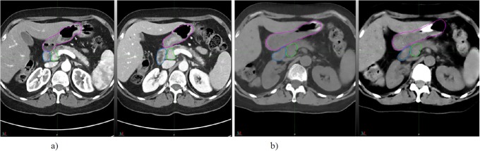
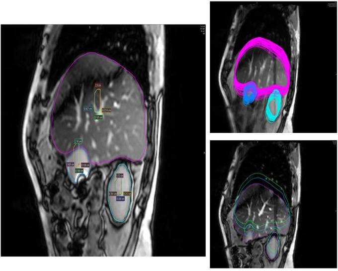
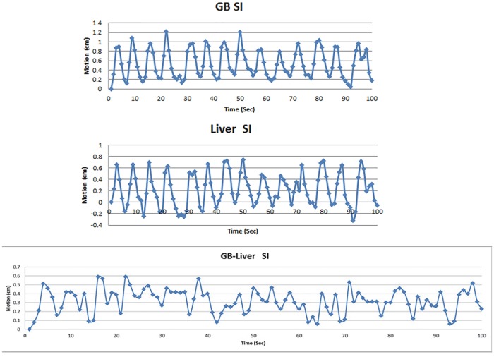
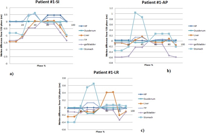
Methods: A series of CT and MRI images acquired with various techniques under free breathing and/or breath hold from 37 randomly-selected pancreatic or liver cancer patients were analyzed to assess the motion in various time frames. These data include FB 4DCT from 15 patients (for motion in time duration of 5 sec), FB 2D cine-MRI from 4 patients (time duration of 1.7 min, 1 second acquisition time per slice), FB cine-MRI acquired using MR-Linac from 6 patients in various fractions (acquisition time is less than 0.6 seconds per slice), FB 4DMRI from 2 patients (time duration of 2 min), respiration-gated T2 with gating at the end expiration (time duration of 3-5 min), and BH T1 with multiphase dynamic contrast in acquisition times of 17 seconds for each of five phases (pre-contrast, arterial, venous, portal venous and delayed post-contrast) from 10 patients. Motions of various organs including gallbladder (GB) and liver were measured based on these MRI data. The GB motion includes both respiration and peristalsis, while liver motion is primarily respiration. By subtracting liver motion (respiration) from GB motion (respiration and peristalsis), the peristaltic motion, along with small residual motion, was obtained.
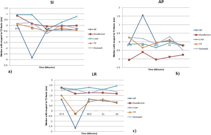
Results: From cine-MRI, the residual motion beyond the respiratory motion was found to be up to 0.6 cm in superior-inferior (SI) and 0.55 cm in anterior-posterior (AP) directions. From 2D cine-MRI acquired by the MR-Linac, different peristaltic motions were found from different fractions for each patient. The peristaltic motion was found to vary between 0.3-1 cm. From BH T1 phase images, the average motions that were primarily due to peristalsis movements were found to be 1.2 cm in SI, 0.7 cm in AP, and 0.9 cm in left-right (LR) directions. The average motions assessed from 4DCT were 1.0 cm in SI and 0.3 cm in AP directions, which were generally smaller than the motions assessed from cine-MRI, i.e., 1.8 cm in SI and 0.6 cm in AP directions, for the same patients. However, average motions from 4DMRI, which are coming from respiratory were measured to be 1.5, 0.5, and 0.4 cm in SI, AP, and LR directions, respectively.
Conclusion: The abdominal motion due to peristalsis can be similar in magnitude to respiratory motion as assessed. These motions can be irregular and persistent throughout the imaging and RT delivery procedures, and should be considered together with respiratory motion during RT for abdominal tumors.
Conflict of interest statement
The authors have no competing interests, although this work was partially funded by Elekta Inc. This does not alter our adherence to PLOS ONE policies on sharing data and materials.
Figures
References
-
- Goldstein SD, Ford EC, Duhon M, McNutt T., Wong J., Herman J. M., Use of respritorycorrelated four-dimensional computed tomography to determine acceptable treatment mergins for locally advanced pancreatic. International Journal of Radiation Oncology Biology Physics. 2010;76: 597–602. - PubMed
-
- Jayachandran P, Minn AY, Van Dam J, Norton J.A., Koong A.C., Chang D.T., Interfractional uncertainty in the treatment of pancreatic cancer with radiation. International Journal of Radiation Oncology Biology Physics. 2010; 76: 603–607. - PubMed
-
- Li XA, Stepaniak C, Gore E. Technical and dosimetric aspects of respiratory gating using a pressure-sensor motion monitoring system. Medical Physics. 2006; 33:145–154. - PubMed
-
- Li X.A., Qi X. S., Pitterle M., Kalakota K., Mueller K., Erickson B. A., et al., “Interfractional variations in patient setup and anatomic change assessed by daily computed tomography” International Journal of Radiation Oncology Biology Physics; 2007; 68: 581–591. - PubMed
Publication types
MeSH terms
LinkOut - more resources
Full Text Sources
Medical
Molecular Biology Databases
