. 2018 Dec:125:140-148.
doi: 10.1016/j.yjmcc.2018.10.009. Epub 2018 Oct 22.

N-terminal extension in cardiac myosin-binding protein C regulates myofilament binding

Affiliations

N-terminal extension in cardiac myosin-binding protein C regulates myofilament binding

Thomas A Bunch et al. J Mol Cell Cardiol. 2018 Dec.

Abstract

Rationale: Mutations in the gene encoding the sarcomeric protein cardiac myosin-binding protein C (cMyBP-C) are a leading cause of hypertrophic cardiomyopathy (HCM). Mouse models targeting cMyBP-C and use of recombinant proteins have been effective in studying its roles in contractile function and disease. Surprisingly, while the N-terminus of cMyBP-C is important to regulate myofilament binding and contains many HCM mutations, an incorrect sequence, lacking the N-terminal 8 amino acids has been used in many studies.

Objectives: To determine the N-terminal cMyBP-C sequences in ventricles and investigate the roles of species-specific differences in cMyBP-C on myofilament binding.

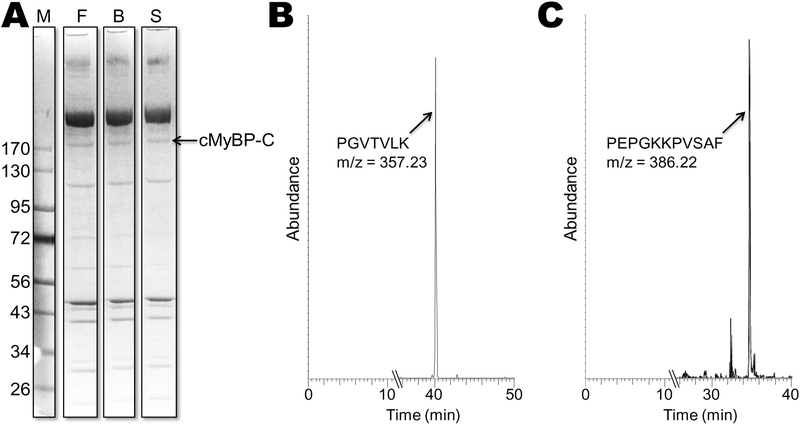
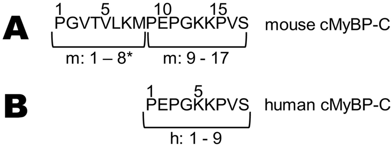
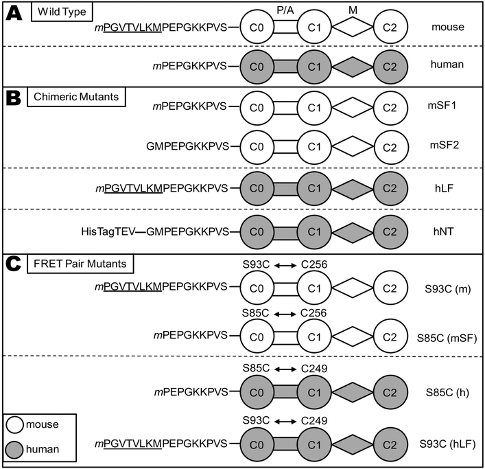
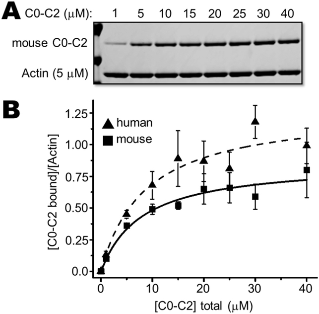
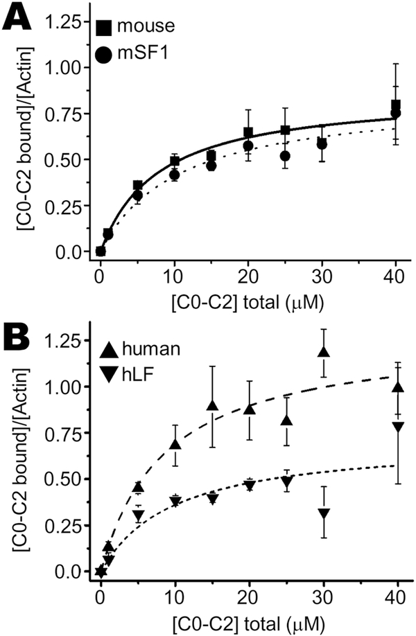
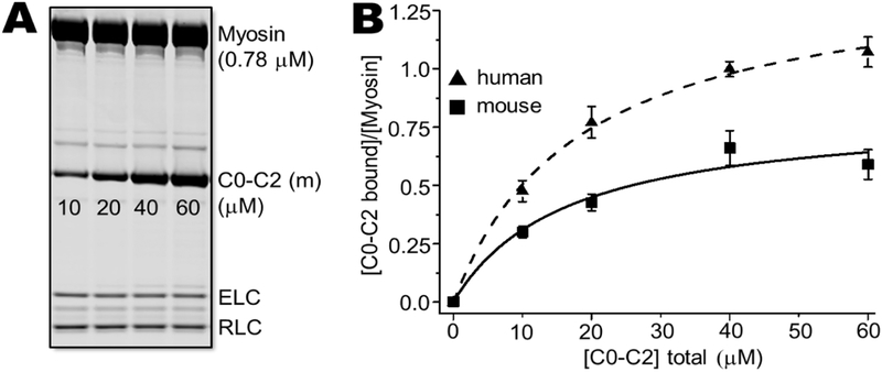
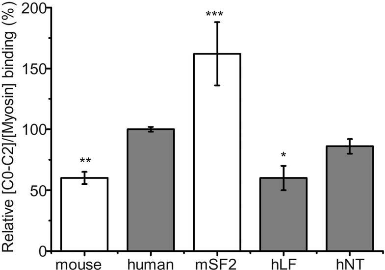
Methods and results: We determined cMyBP-C sequences in mouse and human by inspecting available sequence databases. N-terminal differences were confirmed using liquid chromatography-tandem mass spectrometry (LC-MS/MS). Cosedimentation assays with actin or myosin were used to examine binding in mouse, human and chimeric fusion proteins of cMyBP-C. Time-resolved FRET (TR-FRET) with site-directed probes on cMyBP-C was employed to measure structural dynamics. LC-MS/MS supported the sequencing data that mouse cMyBP-C contains an eight-residue N-terminal extension (NTE) not found in human. Cosedimentation assays revealed that cardiac myosin binding was strongly influenced by the presence of the NTE, which reduced binding by 60%. 75% more human C0-C2 than mouse bound to myosin. Actin binding of mouse C0-C2 was not affected by the NTE. 50% more human C0-C2 than mouse bound to actin. TR-FRET indicates that the NTE did not significantly affect structural dynamics across domains C0 and C1.

Conclusions: Our functional results are consistent with the idea that cardiac myosin binding of N-terminal cMyBP-C is reduced in the mouse protein due to the presence of the NTE, which is proposed to interfere with myosin regulatory light chain (RLC) binding. The NTE is a critical component of mouse cMyBP-C, and should be considered in extrapolation of studies to cMyBP-C and HCM mechanisms in human.

Keywords: Cardiac myosin-binding protein C; Contractile proteins; Myofilament; Myosin; N-terminal extension; Regulation.


Conflict of interest statement

Disclosures: Nothing to disclose.

Figures

Fig. 1.
Fig. 1.. Mass Spectrometry.
(A) Myofibrillar proteins from ventricle isolated from 3 mouse inbred strains (F, FVB/N; B, Black 6; and S, SVE129) separated by SDS-PAGE. The single cMyBP-C band was identified as running ~175 kD was prepared for LC-MS/MS analysis. (B) LC-MS/MS resulting total ion chromatogram of the trypsin peptide from the cMyBP-C band from one of the strains. This peptide peak (PGVTVLK; m/z = 357.23) was present in all 3 strains. (C) Ion chromatogram of the α-chymotrypsin peptide peak (PEPGKKPVSAF; m/z = 386.22) from cMyBP-C from a human ventricular sample.
Fig. 2.
Fig. 2.. N-terminal sequences.
Amino acid sequences of (A) mouse and (B) human cMyBP-C N-termini prior to C0 (see PDB: 2K1M). Methionine residues were not present as starting residues in either group due to aminopeptidase-catalyzed methionine removal. Residues 1–8 of mouse cMyBP-C are the N-terminal extension (NTE)*.
Fig. 3.
Fig. 3.. C0-C2 constructs.
Schematic representation of the cMyBP-C “C0-C2” fragment constructs used in this study. (A) Wild Type C0-C2 constructs differ in their N-terminal sequence, with the mouse (white) isoform containing an N-terminal extension (NTE, underlined) relative to human (gray). All constructs, with the exception of mSF2 and hNT, (defined below) contain a TEV protease-cleavable C-terminal 6× His tag (extending from domain C2) for purification that is removed for functional and structural studies, (cleaved tags not shown). Starting methionine residues (m) were not present in the mature proteins. (B) Mouse short form (mSF1) has the 8-residue NTE removed from the N-terminus and used for actin binding and TR-FRET studies. A second mouse short form (mSF2) was constructed as an N-terminal tagged protein and the resulting protein after removal of the tag with TEV protease begins GMPEP (see methods) and used for myosin binding studies. Human long form (hLF) has the 8-residue mouse NTE added to the N-terminus. Human N-terminal tagged (hNT) C0-C2, used as an uncleaved control for myosin binding assays to non-specifically extend the N-terminus, contains a 6× His tag, 15 amino acids from the pET45 vector, and the TEV recognition sequence for a total of 28 amino acids at the amino terminus prior to GMPEP. (C) WT C0-C2 has a single endogenous Cys in domain C1 (C249 human, C256 mouse) that is readily labeled by thiol-reactive probes. A second Cys in C0 is introduced by site-directed mutagenesis (S85C in human; S93C in mouse) in order to measure FRET distances from C0 to C1. The S93C/S85C were also introduced into the mSF1/hLF chimeras for TR-FRET.
Fig. 4.
Fig. 4.. Actin cosedimentation assays: mouse versus human C0-C2.
Mouse cMyBP-C fragment of C0-C2 with its wild type NTE binding to actin filaments was determined by cosedimentation. (A) Representative high-speed cosedimentation binding experiment with increasing amounts of mouse C0-C2 and constant 5 μM actin. (B) Binding curves of wild type mouse and human C0-C2 fit to a quadratic equation (see Methods). Each experiment was done with independently prepared C0-C2 proteins (n = 4–6). See Table 1 for summary of actin binding results for apparent Kd and Bmax values.
Fig. 5.
Fig. 5.. Actin cosedimentation assays: NTE chimeric mutants.
Chimeric NTE mutant cMyBP-C fragments of C0-C2 compared to wild type mouse or human C0-C2 binding to actin filaments was determined by cosedimentation. Cosedimentation binding experiment with increasing amounts of (A) mouse WT or mSF1 C0-C2, or (B) human WT or hLF C0-C2, and constant 5 μM actin. Binding curves fit to a quadratic equation (see Methods). Each experiment was done with independently prepared C0-C2 proteins (n = 4–6). See Table 1 for summary of actin binding results for chimeric C0-C2.
Fig. 6.
Fig. 6.. Myosin cosedimentation assays: mouse versus human C0-C2.
Human and mouse C0-C2 (10–60 μM) were incubated with 0.78 μM cardiac myosin. Myosin filaments and bound C0-C2 were cosedimented and analyzed by SDS-PAGE. (A) SDS-PAGE gel showing for human C0-C2 binding. (B) Binding curves fit to a quadratic for mouse (squares) and human (triangles) C0-C2 binding to myosin. Each experiment was done with independently prepared C0-C2 proteins (n = 6).
Fig. 7.
Fig. 7.. Myosin cosedimentation assays: NTE chimeric mutants.
Myosin cosedimentation assays comparing mouse (white), human (gray) and N-terminal variant C0-C2 proteins of short form (no NTE) mouse C0-C2 (mSF2), long form (with the mouse NTE) human C0-C2 (hLF), and N-terminal tagged human C0-C2 (hNT) control. Mole fraction (relative to myosin) binding was determined for all forms at 40 μM C0-C2 and 0.78 μM mouse cardiac myosin. Values were normalized to the level of cosedimentation observed for human C0-C2 (see Methods). *p < 0.01, **p < 0.005, and ***p < 0.0001 indicate a significant difference as compared to wild type human C0-C2.

References

    1. Semsarian C, Ingles J, Maron MS, Maron BJ, New perspectives on the prevalence of hypertrophic cardiomyopathy, J Am Coll Cardiol 65(12) (2015) 1249–54. - PubMed
    1. Gersh BJ, Maron BJ, Bonow RO, Dearani JA, Fifer MA, Link MS, Naidu SS, Nishimura RA, Ommen SR, Rakowski H, Seidman CE, Towbin JA, Udelson JE, Yancy CW, G. American College of Cardiology Foundation/American Heart Association Task Force on Practice, 2011 ACCF/AHA Guideline for the Diagnosis and Treatment of Hypertrophic Cardiomyopathy: a report of the American College of Cardiology Foundation/American Heart Association Task Force on Practice Guidelines. Developed in collaboration with the American Association for Thoracic Surgery, American Society of Echocardiography, American Society of Nuclear Cardiology, Heart Failure Society of America, Heart Rhythm Society, Society for Cardiovascular Angiography and Interventions, and Society of Thoracic Surgeons, J Am Coll Cardiol 58(25) (2011) e212–60. - PubMed
    1. Theis JL, Bos JM, Theis JD, Miller DV, Dearani JA, Schaff HV, Gersh BJ, Ommen SR, Moss RL, Ackerman MJ, Expression patterns of cardiac myofilament proteins: genomic and protein analysis of surgical myectomy tissue from patients with obstructive hypertrophic cardiomyopathy, Circ Heart Fail 2(4) (2009) 325–33. - PMC - PubMed
    1. Xu Q, Dewey S, Nguyen S, Gomes AV, Malignant and benign mutations in familial cardiomyopathies: insights into mutations linked to complex cardiovascular phenotypes, J Mol Cell Cardiol 48(5) (2010) 899–909. - PubMed
    1. Tardiff JC, Carrier L, Bers DM, Poggesi C, Ferrantini C, Coppini R, Maier LS, Ashrafian H, Huke S, van der Velden J, Targets for therapy in sarcomeric cardiomyopathies, Cardiovasc Res 105(4) (2015) 457–70. - PMC - PubMed

Publication types