Review
. 2018 Oct 24;100(2):276-293.
doi: 10.1016/j.neuron.2018.09.040.

Towards an Understanding of Synapse Formation

Affiliations
Review

Towards an Understanding of Synapse Formation

Thomas C Südhof. Neuron. .

Abstract

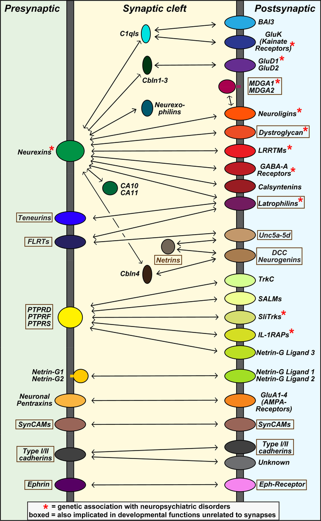
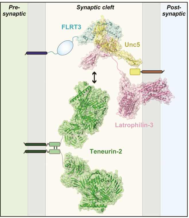
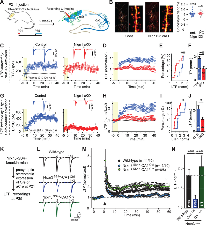
Synapses are intercellular junctions specialized for fast, point-to-point information transfer from a presynaptic neuron to a postsynaptic cell. At a synapse, a presynaptic terminal secretes neurotransmitters via a canonical release machinery, while a postsynaptic specialization senses neurotransmitters via diverse receptors. Synaptic junctions are likely organized by trans-synaptic cell-adhesion molecules (CAMs) that bidirectionally orchestrate synapse formation, restructuring, and elimination. Many candidate synaptic CAMs were described, but which CAMs are central actors and which are bystanders remains unclear. Moreover, multiple genes encoding synaptic CAMs were linked to neuropsychiatric disorders, but the mechanisms involved are unresolved. Here, I propose that engagement of multifarious synaptic CAMs produces parallel trans-synaptic signals that mediate the establishment, organization, and plasticity of synapses, thereby controlling information processing by neural circuits. Among others, this hypothesis implies that synapse formation can be understood in terms of inter- and intracellular signaling, and that neuropsychiatric disorders involve an impairment in such signaling.

Keywords: BAIs; cell-adhesion molecules; cerebellins; latrophilins; neurexins; neuroligins; synapse; synaptic plasticity; synaptogenesis; teneurins.


Figures

Figure 1:
Figure 1:. Canonical design of a central synapse
Schematic drawing of a synaptic junction with a cluster of synaptic vesicles (SV) on the presynaptic side, and an array of neurotransmitter receptors on the postsynaptic side. Pre- and postsynaptic transport vesicles for receptors, active zone components, and transsynaptic cell adhesion molecules are indicated, as well as presynaptic endosomes and postsynaptic organelles (Golgi apparatus, endosomes, and endoplasmic reticulum [ER]). Note that in brain, the synaptic cleft is usually wider than the surrounding interstitial space. All synapses contain similar presynaptic components independent of type, although the specific isoforms of various proteins (synaptotagmins, neurotransmitter transporters, RIMs and Munc13, Ca2+-channels etc) vary. In contrast, postsynaptic components of excitatory and inhibitory synapses exhibit no homology, neither at the level of receptors nor in the postsynaptic scaffolding proteins. Moreover, presynaptic specializations are formed exclusively by neurons, but postsynaptic specializations can likely be formed by any cell in the body.
Figure 2:
Figure 2:. Flow diagram of synapse formation
During development, neurons are generated, migrate, and grow short and long-range axons and extensive dendritic trees. Axons and dendrites establish initial synaptic contacts mostly during development and the early postnatal period, although synapse formation continues throughout life. Synapse formation is represented as a multicomponent process whereby an initial synaptic contact nucleates organization of pre- and postsynaptic specializations that are subsequently specified, i.e., become endowed with specific properties. Synapse specification is likely an activity-dependent process that takes place continuously for most synapses in brain as synapse are being restructured during synaptic plasticity. Most physiological synapse elimination occurs during the first decades of life, but a low level of synapse elimination, like synapse formation, continues throughout life. Pathological synapse elimination may be induced by synapse dysfunction and is a hallmark of neurodegenerative disorders (e.g., Alzheimer’s disease).
Figure 3:
Figure 3:. Artificial synapse formation assay
A, Exemplary list of synaptic CAMs that induce either pre- or postsynaptic specializations during artificial synapse formation assays, which involve expression of a candidate synaptic CAM in a non-neuronal cell, such as a HEK293 or COS cell, and co-culture of this non-neuronal cell with dissociated neurons. Synaptic CAMs under these conditions induce formation of either pre- or postsynaptic specializations by contacting neurons, but never both. B, Representative image of presynaptic specializations in co-cultured neurons induced by neuroligin-1 that is expressed in a COS cell. Presynaptic terminals are visualized by staining for synapsin and shown in green, while neuroligin-1 staining is shown in red, and overlapping signals are shown in yellow. Green dots outside of the COS cell surface represent synapses between the co-cultured neurons. C, Representative image of a transmission electron micrograph of a COS cell expressing neuroligin-1 that has been co-cultured with dissociated mouse neurons. Artificial synaptic contacts are extremely abundant and exhibit a normal synaptic morphology, including an apparent postsynaptic density. Note that the size of the synaptic contacts are uniform similar to normal synapses, even though they are formed often by giant nerve terminals as an array of specializations. Panels B and C are modified from Chubykin et al. (2005).
Figure 4:
Figure 4:. Cartoon of candidate trans-synaptic CAM interactions
Summary of prominent CAM interactions that were proposed to operate at the synapse. CAMs were placed on the pre- or postsynaptic side based on the overall published studies, but for many molecules firm assignments cannot yet be made. Some of the interactions shown are supported by compelling biophysical evidence (e.g., binding of LAR-type RPTPs to their various ligands), but others are more tenuous (e.g., binding of neurexins to latrophilins or to C1ql’s). Diagram was modified from Südhof (2017).
Figure 5:
Figure 5:. Atomic structure of the trans-synaptic teneurin-latrophilin-FLRT complex
Atomic structures determined for FLRT3 (blue), Unc5 (yellow), Lphn3 (pink), and teneurin2 (green) are placed into synaptic cleft of an excitatory synapse. Teneurin-2 is positioned presynaptically because it forms a trans-cellular junction with postsynaptic latrophilins (Boucard et al., 2014) and because it induces postsynaptic specializations in the artificial synapse formation assay (Li et al., 2018). FLRT3 is also positioned presynaptically because it forms trans-cellular complexes with Lphn2 (Lu et al., 2015). Note that latrophilins were originally thought to be presynaptic and FLRTs postsynaptic based on RNAi data (O’Sullivan et al., 2012), but conditional genetic knockouts established that in cultured neurons and in vivo, at least Lphn2 is exclusively postsynaptic (Anderson et al., 2016). Interestingly, presynaptic FLRT3 can bind simultaneously to both postsynaptic Lphn3 and postsynaptic Unc5, as shown by crystallography of the complex (Lu et al., 2015). It is unknown whether Lphn3 can simultaneously bind to FLRT3 and to teneurins, or whether these interactions are mutually exclusive, but in either case the various binding reactions suggest that a trans-synaptic interaction network anchored on latrophilins and teneurins may form the basis for an extensive signaling machinery at the synapse. Structures are from Lu et al. (2015) for the FLRT3/Unc5/Lphn3 complex; Arac et al. (2011) for the Lphn3 GAIN/hormone binding domain fragment; and Li et al. (2018) for teneurin (PDB IDs: teneurin-2, 6CMX; Lphn3, 4DLQ (GAIN and hormone-binding domains), 5CMN/5AFB (lectin and olfactomedin domains) and 4K5Y (transmembrane GPCR domain of the corticotrophin releasing hormone receptor); FLRT, 5CMN; and Unc5, 5FTT). The figure was prepared by Drs. Demet Arac and Jingxian Li (U. of Chicago).
Figure 6:
Figure 6:. Illustration of experiments that probe the role of trans-synaptic CAM interactions involving postsynaptic neuroligin-1 (Nlgn1) and presynaptic neurexin-3 (Nrxn3) in NMDA-receptor dependent LTP as a synapse restructuring event
A, Experimental paradigm. The hippocampus of conditionally mutant mice are infected at P21 by stereotactic injections with viruses expressing inactive (ΔCre, control) or active Cre-recombinase (Cre), and analyzed after 2–3 weeks by acute slice physiology. B, Deletion of all major neuroligins does not change spine density. Hippocampal CA1 region neurons of triple conditional knockout (cKO) mice in which all three major neuroligin genes (Nlgn1-Nlgn3) are floxed were sparsely infected with lentiviruses, and the spine density as a proxy for synapse density was examined (left, sample images; right, summary graph). C-F, Conditional neuroligin-1 (Nlgn1) deletion blocks NMDAR-dependent LTP (C, exemplary experiments with sample traces above; D, average data; E, cumulative plot of LPT as a function of cell number; E, summary graph of the extent of LTP). G-J, Same as C-F, but for LTP induced by prolonged postsynaptic depolarization in the presence of the NMDA-receptor blocker AP5. K-N, Constitutive inclusion of splice site #4 (SS4) in presynaptic neurexin-3 (Nrxn3) transsynaptically blocks postsynaptic LTP; this block can be reversed by presynaptic conversion of SS4+ to SS4- Nrxn3 (K, experimental strategy; L, sample traces; M, summary plots of LTP monitored in slices from wild-type control mice and from Nrxn3SS#4+ knockin mice in which all neurons express only the SS#4+ variants of Nrxn3 isoforms and which were presynaptically injected with AAVs encoding ΔCre (to retain Nrxn3-SS#4+ expression) or Cre (to convert presynaptic Nrxn3-SS#4+ into Nrxn3-SS#4-; N, summary graph of the extent of LTP). Data in B-J and K-N are modified from Jiang et al. (2016) and Aoto et al. (2013), respectively.

References

    1. Araç D, Boucard AA, Bolliger MF, Nguyen J, Soltis M, Südhof TC, Brunger AT (2011) A Novel Evolutionarily Conserved Domain of Cell-Adhesion GPCRs Mediates Autoproteolysis. EMBO J. 31, 1364–1378. - PMC - PubMed
    1. Ackley BD, Harrington RJ, Hudson ML, Williams L, Kenyon CJ, Chisholm AD, Jin Y. (2005) The two isoforms of the Caenorhabditis elegans leukocyte-common antigen related receptor tyrosine phosphatase PTP-3 function independently in axon guidance and synapse formation. J. Neurosci. 25, 7517–7528. - PMC - PubMed
    1. Acuna C, Liu X, Südhof TC (2016) How to Make an Active Zone: Unexpected Universal Functional Redundance between RIMs and RIM-BPs. Neuron 91, 792–807. - PubMed
    1. Aoto J, Martinelli DC, Malenka RC, Tabuchi K, Südhof TC (2013) Presynaptic Neurexin-3 Alternative Splicing Trans-Synaptically Controls Postsynaptic AMPA-Receptor Trafficking. Cell 154, 75–88. - PMC - PubMed
    1. Araki Y, Zeng M, Zhang M, Huganir RL (2015) Rapid dispersion of SynGAP from synaptic spines triggers AMPA receptor insertion and spineenlargement during LTP. Neuron 85, 173189. - PMC - PubMed

Publication types

Substances

LinkOut - more resources