Review
. 2018 Oct 24;100(2):294-313.
doi: 10.1016/j.neuron.2018.10.009.

Development and Functional Diversification of Cortical Interneurons

Affiliations
Review

Development and Functional Diversification of Cortical Interneurons

Lynette Lim et al. Neuron. .

Abstract

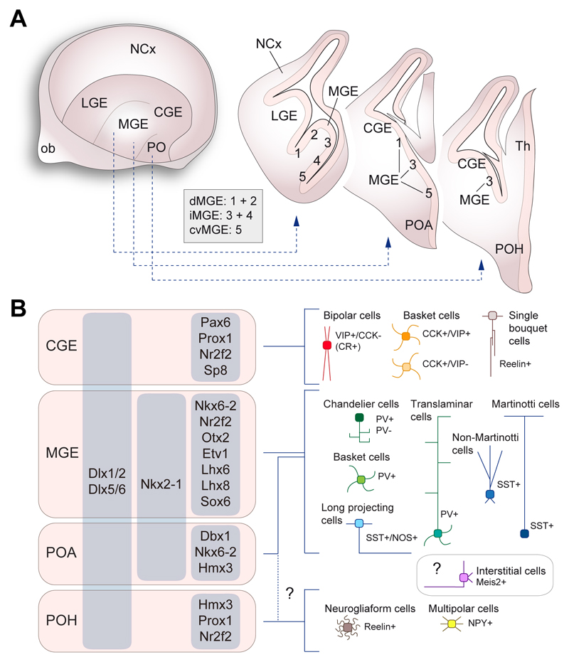
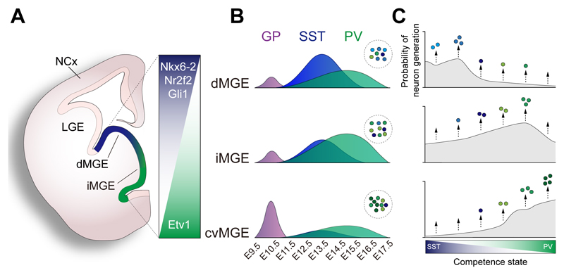
In the cerebral cortex, GABAergic interneurons have evolved as a highly heterogeneous collection of cell types that are characterized by their unique spatial and temporal capabilities to influence neuronal circuits. Current estimates suggest that up to 50 different types of GABAergic neurons may populate the cerebral cortex, all derived from progenitor cells in the subpallium, the ventral aspect of the embryonic telencephalon. In this review, we provide an overview of the mechanisms underlying the generation of the distinct types of interneurons and their integration in cortical circuits. Interneuron diversity seems to emerge through the implementation of cell-intrinsic genetic programs in progenitor cells, which unfold over a protracted period of time until interneurons acquire mature characteristics. The developmental trajectory of interneurons is also modulated by activity-dependent, non-cell-autonomous mechanisms that influence their ability to integrate in nascent circuits and sculpt their final distribution in the adult cerebral cortex.

Keywords: GABA; cerebral cortex; inhibitory; interneuron; migration; neocortex; neural circuit; neuronal identity; specification; transcription factor.


Figures

Figure 1
Figure 1. Milestones in the development of cortical interneurons
(A) Timeline of the development of cortical interneurons in the mouse. The main events have been highlighted in corresponding temporal periods: neurogenesis, tangential migration, laminar allocation (which involves radial migration), wiring (dendritic and axonal morphogenesis and establishment of synapses), programmed cell death and circuit refinement. Interneuron identity is specified at neuronal birth, but it unfolds over a protracted period of time through which the final characteristics of each type of interneuron are acquired. (B) The development of layer 2/3 SST+ Martinotti cells is used here as an example to illustrate the main developmental milestones in the generation of cortical interneurons in mice. At least a population of SST+ Martinotti cells is generated from progenitor cells in the dorsal aspect of the MGE. SST+ Martinotti cells preferentially migrate to the embryonic cortex through the marginal zone (MZ) stream. During radial migration into the cortical plate (CP), SST+ Martinotti cells leave their trailing neurite in the MZ, which will eventually develop into a characteristic axonal arborization in layer 1. By the end of the first postnatal week, about 30% of interneurons undergo program cell death, including SST+ Martinotti cells. This process depends on the integration of these cells into cortical circuits. The surviving SST+ Martinotti cells remodel their synaptic connections during the second and third week of postnatal development. For example, layer 2/3 SST+ Martinotti cells end up establishing preferential connections with the apical dendrites of pyramidal cells also located in layer 2/3. The yellow thunderbolt symbol indicates processes that depend on neuronal activity. MGE, medial ganglionic eminence; NCx, neocortex; SVZ, subventricular zone; VZ, ventricular zone.
Figure 2
Figure 2. Modular organization of cortical interneuron features
(A) Cortical interneurons are characterized by a combination of morphological, biochemical, intrinsic and connectivity properties. The most prominent morphological characteristics of cortical interneurons are the shape and orientation of dendrites and axons. Biochemical properties define important aspects of synaptic communication, including the co-release of neuropeptides along with GABA. Intrinsic electrophysiological properties are largely determined by the combination of ionic channels they express. Interneurons establish synapses on different subcellular compartments of their targets, which impact their role in neural circuits. (B) The combinatorial organization of cortical interneurons is illustrated for PV+ basket cells (green) and CCK+ basket cells (orange). Both cell types share similar morphology and connectivity principles, but they differ in their intrinsic and biochemical properties. (C) The schema depicts how each set of defining features might be acquired through a combination of partially shared transcriptional programs.
Figure 3
Figure 3. Diversity of GABAergic neurons in the neocortex
(A) Schemas depicting the main classes of cortical interneuron in the mouse neocortex. Interneurons can be organized into three large classes based on the expression of PV, SST and the serotonin receptor 3A (Htr3a). A small fraction of PV+ basket cells also express SST. (B) Schematic depicting the general laminar distribution of interneuron types in the neocortex. Some cell types are found in most layers of the cortex (e.g., PV+ basket cells), whereas others seem to have a much more restricted laminar distribution (e.g., Meis2+ cells). (C) The schematic illustrates the approximate relative frequency of each type of interneuron, color-coded as in (A). It should be noted here are no direct estimates of the frequency of chandelier cells and many other classes of interneuron. In addition, the relative proportion of interneurons likely varies across different cortical areas. cc, corpus callosum; wm, white matter.
Figure 4
Figure 4. Developmental origin of cortical interneurons
(A) Regional organization of the embryonic subpallium. The schematic on the left depicts a telencephalic hemisphere viewed from the midline and from which medial structures have been removed. The drawings on the right illustrate the organization of the subpallium in three coronal sections through the telencephalon. The medial ganglionic eminence (MGE) consists of several progenitor domains (numbered 1 to 5) as described by Flames and colleagues (2007) and can be broadly subdivided in dorsal (dMGE), intermediate (iMGE) and caudoventral (cvMGE) regions. (B) Regional origin of the main types of cortical interneurons. The preoptic region (PO) comprise two progenitor domains, the preoptic area (POA) and the preoptic-hypothalamic (POH) border domain. The precise origin of neurogliaform and multipolar cells within these domains remain uncertain. The origin of interstitial Meis2+ cells also remains unclear. CGE, caudal ganglionic eminence; LGE, lateral ganglionic eminence; NCx, neocortex, PO, preoptic region.
Figure 5
Figure 5. Spatiotemporal patterning of interneuron neurogenesis
(A) Schematic representation of a coronal section through the anterior aspect of the medial ganglionic eminence (MGE). Several transcription factors contribute to the spatial patterning of MGE into different domains: dorsal (dMGE), intermediate (iMGE) and caudoventral (cvMGE). (B) Spatio-temporal specification of cortical interneurons. Progenitor pools within the MGE generate different numbers of neurons following a temporal sequence. All domains generate an early cohort of projection neurons for the globus pallidus (GP), followed by SST+ and PV+ interneurons in different ratios. The area under each curve represents relative numbers of neurons arising from each MGE domain. (C) An integrated model of interneuron generation. In this model, MGE progenitors undergo progressive intrinsic changes in their competence to generate different types of neuron during development. For any MGE progenitor, the generation of more or less neurons of a specific class is determined by the probability to generate cells during a given competence state. Color gradients represent progression of a single progenitor cells across different competency states, while the curves represent the probability of cell generation in each state. Progenitors in each MGE region would have different probabilistic rules, which would lead to the differences in the relative number of interneurons of each type generated in each region.
Figure 6
Figure 6. Activity dependent regulation of interneuron assembly
(A) Postnatal maturation and assembly of interneurons into cortical circuits. During the first postnatal week, cortical interneurons invade the cortical plate and begin to interact with surrounding pyramidal neurons as well as other local interneurons and thalamic afferents. At early postnatal stages (P0 to P5) these interactions are required for the correct morphological maturation of most types of interneuron. Following that period, about a third of cortical interneurons undergo programmed cell death. Activity-dependent mechanisms are critical for the survival of the remaining cortical interneurons. (B) Activity dependent regulation of interneuron numbers. Cortical interneurons are programmed to die in the absence of external signals, most of which are likely provided by pyramidal cells. Consistently, reduction of interneuron or pyramidal cell activity decreases interneuron survival, while increase in the number or activity of excitatory neurons promotes the survival of a larger fraction of interneurons.
Figure 7
Figure 7. Dynamic regulation of interneuron properties
(A) The expression of the transcription factor Npas4 is regulated by activity in SST+ interneurons. Increased network activity, for example through sensory stimulation, leads to increased levels of Npas4, which in turn regulates the transcription of genes encoding proteins that increase the number of excitatory synapses SST+ interneurons receive. (B) Activity negatively regulates the expression of the transcription factor Etv1 and the perineuronal net protein Brevican (Bcan) in PV+ basket cells. Etv1 regulates the expression of genes encoding proteins that regulate the intrinsic properties of PV+ basket cells (e.g., Kv1.1) and the number of excitatory synapses these interneurons receive. Bcan levels are also linked to the expression of proteins regulating the intrinsic properties of PV+ basket cells (e.g., Kv3.1) and their synaptic inputs (e.g., AMPAR).
None

References

    1. Anastasiades PG, Marques-Smith A, Lyngholm D, Lickiss T, Raffiq S, Katzel D, Miesenbock G, Butt SJ. GABAergic interneurons form transient layer-specific circuits in early postnatal neocortex. Nat Commun. 2016;7 10584. - PMC - PubMed
    1. Anderson SA, Eisenstat DD, Shi L, Rubenstein JLR. Interneuron migration from basal forebrain to neocortex: dependence on Dlx genes. Science. 1997a;278:474–476. - PubMed
    1. Anderson SA, Qiu M, Bulfone A, Eisenstat DD, Meneses J, Pedersen R, Rubenstein JLR. Mutations of the homeobox genes Dlx-1 and Dlx-2 disrupt the striatal subventricular zone and differentiation of late born striatal neurons. Neuron. 1997b;19:27–37. - PubMed
    1. Antypa M, Faux C, Eichele G, Parnavelas JG, Andrews WD. Differential gene expression in migratory streams of cortical interneurons. Eur J Neurosci. 2011;34:1584–1594. - PMC - PubMed
    1. Arshad A, Vose LR, Vinukonda G, Hu F, Yoshikawa K, Csiszar A, Brumberg JC, Ballabh P. Extended production of cortical interneurons into the third trimester of human gestation. Cereb Cortex. 2016;26:2242–2256. - PMC - PubMed

Publication types

LinkOut - more resources