Review
. 2018 Oct 24;100(2):375-388.
doi: 10.1016/j.neuron.2018.09.022.

The Meningeal Lymphatic System: A New Player in Neurophysiology

Affiliations
Review

The Meningeal Lymphatic System: A New Player in Neurophysiology

Sandro Da Mesquita et al. Neuron. .

Abstract

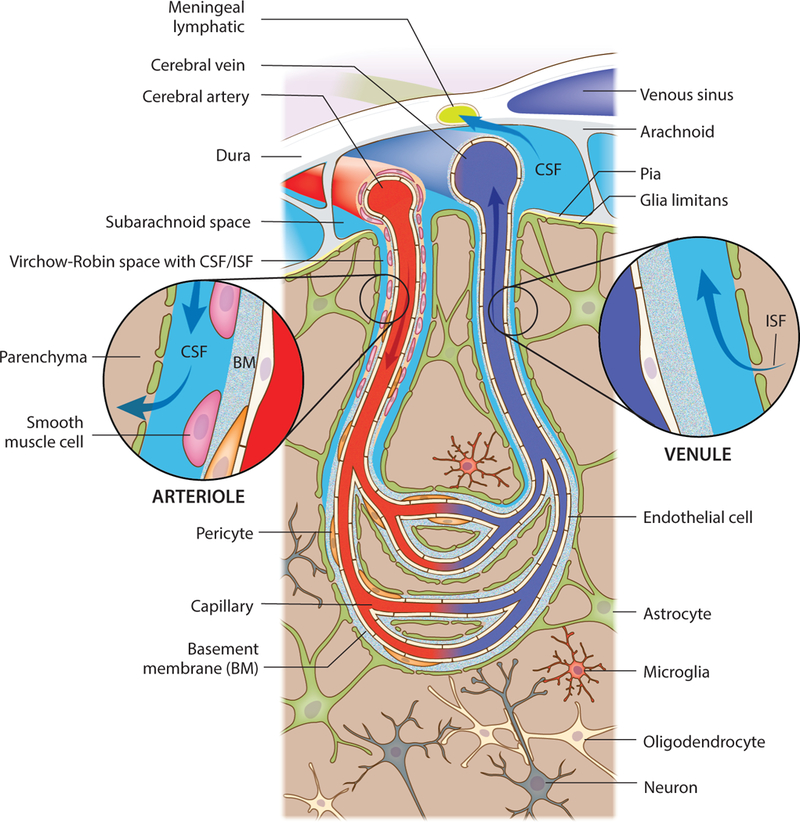
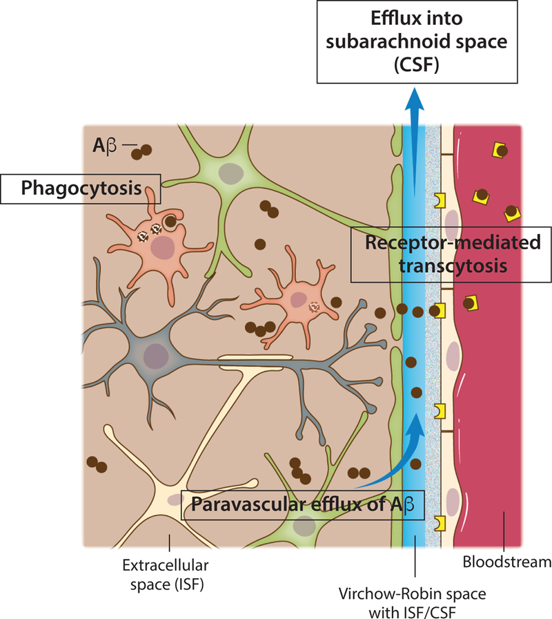
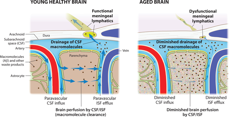
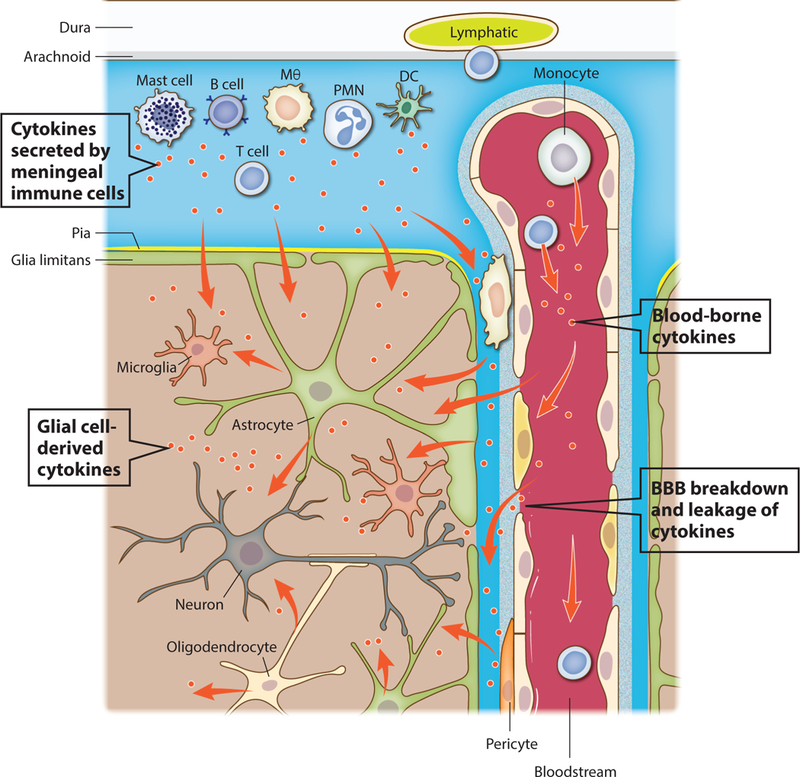
The nature of fluid dynamics within the brain parenchyma is a focus of intensive research. Of particular relevance is its participation in diseases associated with protein accumulation and aggregation in the brain, such as Alzheimer's disease (AD). The meningeal lymphatic vessels have recently been recognized as an important player in the complex circulation and exchange of soluble contents between the cerebrospinal fluid (CSF) and the interstitial fluid (ISF). In aging mammals, for example, impaired functioning of the meningeal lymphatic vessels can lead to accelerated accumulation of toxic amyloid beta protein in the brain parenchyma, thus aggravating AD-related pathology. Given that meningeal lymphatic vessels are functionally linked to paravascular influx/efflux of the CSF/ISF, and in light of recent findings that certain cytokines, classically perceived as immune molecules, exert neuromodulatory effects, it is reasonable to suggest that the activity of meningeal lymphatics could alter the accessibility of CSF-borne immune neuromodulators to the brain parenchyma, thereby altering their effects on the brain. Accordingly, in this Perspective we propose that the meningeal lymphatic system can be viewed as a novel player in neurophysiology.

Keywords: Alzheimer’s disease; cerebrospinal fluid; cytokines; glia; glymphatic; immune cells; interstitial fluid; lymphatic vessels; meninges; neuromodulation.


Conflict of interest statement

Declaration of interests

J.K. is an Advisor to PureTech Health/Ariya.

Figures

Figure 1.
Figure 1.. Cytoarchitecture of the meninges, brain vasculature and pathways of paravascular recirculation
A schematic representation of the brain meninges constituted by dura, arachnoid and pia layers. Lymphatic vessels that are present in the meningeal dura drain components of the cerebrospinal fluid (CSF) that fills the subarachnoid space. Arising from the brain surface, cerebral arteries extend into pial and then subpial arteries. Higher caliber pial arteries extend into smaller caliber arterioles (both wrapped by smooth muscle cells) that dive into the brain parenchyma. Clearly defined paravascular spaces of about 50–100 nm, the Virchow-Robin spaces, are filled with CSF that flows into deeper brain regions, along the arterioles and capillaries, and diffuses through the glia limitans into the parenchyma. Efflux of interstitial fluid (ISF) happens through paravenous spaces back into the subarachnoid CSF.
Figure 2.
Figure 2.. Mechanisms of amyloid beta clearance from the brain
Toxic amyloid beta (Aβ) peptides are present in the extracellular brain ISF. One of the most important mechanisms of Aβ clearance from the brain extracellular environment is receptormediated transcytosis across the blood-brain barrier. Monomeric or aggregated Aβ can also be internalized and degraded by brain phagocytes, resident microglia or monocyte-derived macrophages that might be recruited and engraft the brain. Efflux of soluble Aβ from the brain ISF back into the subarachnoid CSF sink also takes place through paravascular spaces (glymphatic route).
Figure 3.
Figure 3.. Aging diminishes meningeal lymphatic drainage and paravascular recirculation of CSF
macromolecules Functional meningeal lymphatic vessels drain macromolecules (such as Aβ) from the CSF of the young and healthy brain. Influx of CSF through the paravascular (glymphatic) route leads to brain perfusion by CSF/ISF and paravascular efflux of macromolecules from the parenchymal ISF back into the CSF. Dysfunction of meningeal lymphatic vessels with aging contributes to impairment of influx/efflux mechanisms and to poor recirculation of CSF content.
Figure 4.
Figure 4.. Major sources of cytokines in the CNS
Cytokines can act as neuromodulators in both physiological and pathological conditions and their major sources are depicted. One of the major and best characterized sources of cytokines in the brain are parenchymal cells, particularly glial cells like microglia, astrocytes and oligodendrocytes. Cells found in the vicinity of the blood-brain barrier (BBB), namely perivascular macrophages and pericytes, also secrete cytokines that can reach the brain extracellular milieu. Myeloid and lymphoid immune cells present in the brain meninges produce and secrete cytokines into the CSF, which may then be transported into the brain parenchyma trough the glymphatic route. Finally, blood-borne cytokines might act directly on endothelial cells of the brain vasculature (like type I interferons upon infection), or reach the brain parenchyma in conditions where the BBB is disrupted and leaky.

References

    1. Absinta M, Ha SK, Nair G, Sati P, Luciano NJ, Palisoc M, Louveau A, Zaghloul KA, Pittaluga S, Kipnis J, and Reich DS (2017). Human and nonhuman primate meninges harbor lymphatic vessels that can be visualized noninvasively by MRI. Elife 6. - PMC - PubMed
    1. Agorastos A, Hauger RL, Barkauskas DA, Moeller-Bertram T, Clopton PL, Haji U, Lohr JB, Geracioti TD Jr., Patel PM, Chrousos GP, and Baker DG (2014). Circadian rhythmicity, variability and correlation of interleukin-6 levels in plasma and cerebrospinal fluid of healthy men. Psychoneuroendocrinology 44, 71–82. - PubMed
    1. Alitalo K (2011). The lymphatic vasculature in disease. Nat Med 17, 1371–1380. - PubMed
    1. Alitalo K, Tammela T, and Petrova TV (2005). Lymphangiogenesis in development and human disease. Nature 438, 946–953. - PubMed
    1. Antila S, Karaman S, Nurmi H, Airavaara M, Voutilainen MH, Mathivet T, Chilov D, Li Z, Koppinen T, Park JH, et al. (2017). Development and plasticity of meningeal lymphatic vessels. J Exp Med 214, 3645–3667. - PMC - PubMed

Publication types

LinkOut - more resources