Review
. 2018 Oct 24;100(2):406-423.
doi: 10.1016/j.neuron.2018.10.015.

Lost in Translation: Traversing the Complex Path from Genomics to Therapeutics in Autism Spectrum Disorder

Affiliations
Review

Lost in Translation: Traversing the Complex Path from Genomics to Therapeutics in Autism Spectrum Disorder

Nenad Sestan et al. Neuron. .

Abstract

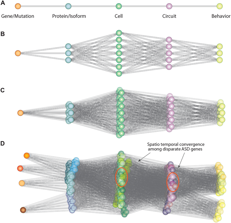
Recent progress in the genomics of non-syndromic autism spectrum disorder (nsASD) highlights rare, large-effect, germline, heterozygous de novo coding mutations. This distinguishes nsASD from later-onset psychiatric disorders where gene discovery efforts have predominantly yielded common alleles of small effect. These differences point to distinctive opportunities for clarifying the neurobiology of nsASD and developing novel treatments. We argue that the path ahead also presents key challenges, including distinguishing human pathophysiology from the potentially pleiotropic neurobiology mediated by established risk genes. We present our view of some of the conceptual limitations of traditional studies of model organisms, suggest a strategy focused on investigating the convergence of multiple nsASD genes, and propose that the detailed characterization of the molecular and cellular landscapes of developing human brain is essential to illuminate disease mechanisms. Finally, we address how recent advances are leading to novel strategies for therapeutics that target various points along the path from genes to behavior.

Keywords: autism spectrum disorder; convergence; convergence neuroscience; de novo mutation; gene therapy; genomics; human brain development; neurodevelopmental disorders; non-syndromic autism spectrum disorder; transcriptomics.


Conflict of interest statement

Declaration of Interests

Matthew W. State serves on the scientific advisory boards and has stock or stock options for BlackThorn therapeutics and ARett pharmaceuticals.

Figures

Figure 1:
Figure 1:. Spatiotemporal convergence among high confidence ASD risk genes
The figure provides a conceptual illustration of the path from risk genes to behavior in nsASD. Panel A illustrates an idealized path from genes to proteins to cells, circuits and behavior. This is shown as a line connecting color spheres representing the noted levels of analysis (orange = cells, blue = proteins; green =cell etc.); Panel B shows the complexity added by consideration of isoform diversity and biological pleiotropy, with a single gene/mutation leading to multiple transcripts and proteins and corresponding to multiple cell types and circuits. Panel C represents the further complexity added by the consideration of spatial and temporal influence on expression and function during brain development. This is shown as a three-dimensional space connecting a single gene to multiple isoforms, protein, cell types and circuits. Sexual dimorphism potentially adds another dimension of complexity. Panel D represents the strategy of using multiple independent risk genes in an effort to triangulate on specific cell types and circuits that show overlap among functionally diverse risk genes.
Figure 2:
Figure 2:. High confidence ASD risk genes encode synaptic proteins and chromatin and transcriptional regulators
Genetic studies have identified a large number of risk genes for ASD, many of which have pleiotropic functional properties. Synapse function, chromatin modification and transcriptional regulation top the list of statistically enriched functional categories. On the left, a simplified schematic of the major cellular components of neural circuits in the cerebral cortex: Pyramid-shaped glutamatergic excitatory projections neurons, GABAergic inhibitory interneurons, and glial cells. On the right, diverse intracellular distribution and pleiotropic roles of ASD risk genes (Sanders et al., 2015). Red circles depict a view of the synapse with its many protein products of ASD risk genes (top) and the ASD proteins in the nucleus (bottom). Proteins in synaptic signaling pathways encompass cell adhesion, scaffolding and signaling molecules. Nuclear protein products of ASD risk genes are mainly associated with chromatin modification and transcriptional control, suggesting that alterations in chromatin structure and gene expression may contribute to ASD.
Figure 3.
Figure 3.. A timeline of major human neurodevelopmental processes and neuropsychiatric disorders
The illustrations in the top panel demonstrate the dramatic changes in the anatomical features of human brain over the course of prenatal and postnatal development. The gray bar provides a timeline of major phases of human development (Kang et al., 2011), which are plotted on a logarithmic scale. Age is shown in postconceptional weeks (PCW) and postnatal years (PY). The second panel shows the age of onset for selected psychiatric and neurological disorders. The beige bars indicate the age range at which each disorder is most commonly diagnosed, with less frequent ages of diagnosis denoted as dotted lines. Note that the age of diagnosis is highly variable between disorders. The schematic in the third, fourth and fifth panel details the approximate timing and sequence of key cellular processes in the prefrontal cortex, the primary visual cortex and cerebellum, respectively. Bars indicate the peak developmental window in which each feature is acquired. Dotted lines indicate that feature acquisition occurs at these ages, though to a relatively minor degree. Arrows indicate that the feature is present throughout postnatal life. DL, deep layer excitatory projection (pyramidal) neurons; ExN, excitatory projection neurons; Granule, granule cells; IN, Interneurons; PN, Purkinje neurons; and UL, upper layer excitatory projection neurons. Based on figures from Silbereis et al., 2016. Light red squares denote two spatiotemporal windows (i.e., the prefrontal and motor-somatosensory cortex during mainly the midfetal development, and thalamus/cerebellar cortex during infancy and early childhood) that were identified in Willsey et al., 2013 to be significantly enriched for risk among high-confidence ASD genes. The shade of red denotes the significance level of the enrichment after correction for multiple comparisons (darker shade denotes greater enrichment). Relevant references pertaining to information detailed in the panels are provided in the rightmost column: a (Kessler et al., 2007) and (Lee et al., 2014), b (Myers, 2004), c (Pagano et al., 2016), d (van der Flier et al., 2011), e (Clancy et al., 2001) (Bystron et al., 2006), f (Paredes et al., 2016), g (Kang et al., 2011); (deAzevedo et al., 2003);(Choi and Lapham, 1978), h (Yeung et al., 2014); (Kang et al., 2011)), i (Petanjek et al., 2011); (Huttenlocher and Dabholkar, 1997);(Kostovic and Rakic, 1990); (Kwan et al., 2012), j (Yakovlev and Lecours 1967); (Gilles, 1976); (Miller et al., 2012), k (Petanjek et al., 2011); (Huttenlocher and Dabholkar, 1997)), l (Clancy et al., 2001)), m (Huttenlocher and Dabholkar, 1997), n (Sidman and Rakic, 1973), o (Kiessling et al., 2014; Riedel et al., 1989), p (Yakovlev and Lecours 1967); (Gilles, 1976)).

References

    1. Autism Genome Project C, Szatmari P, Paterson AD, Zwaigenbaum L, Roberts W, Brian J, Liu XQ, Vincent JB, Skaug JL, Thompson AP, et al. (2007). Mapping autism risk loci using genetic linkage and chromosomal rearrangements. Nat Genet 39, 319–328. - PMC - PubMed
    1. Autism Spectrum Disorders Working Group of The Psychiatric Genomics, C. (2017). Meta-analysis of GWAS of over 16,000 individuals with autism spectrum disorder highlights a novel locus at 10q24.32 and a significant overlap with schizophrenia. Mol Autism 8, 21. - PMC - PubMed
    1. Bae BI, Jayaraman D, and Walsh CA (2015). Genetic changes shaping the human brain. Dev Cell 32, 423–434. - PMC - PubMed
    1. Barak T, Kwan KY, Louvi A, Demirbilek V, Saygi S, Tuysuz B, Choi M, Boyaci H, Doerschner K, Zhu Y, et al. (2011). Recessive LAMC3 mutations cause malformations of occipital cortical development. Nat Genet 43, 590–594. - PMC - PubMed
    1. Bauman MD, Iosif AM, Ashwood P, Braunschweig D, Lee A, Schumann CM, Van de Water J, and Amaral DG (2013). Maternal antibodies from mothers of children with autism alter brain growth and social behavior development in the rhesus monkey. Transl Psychiatry 3, e278. - PMC - PubMed

Publication types