. 2019 Mar;155(3):534-539.
doi: 10.1016/j.chest.2018.10.007. Epub 2018 Oct 22.

Tissue Factor Facilitates Wound Healing in Human Airway Epithelial Cells

Affiliations

Tissue Factor Facilitates Wound Healing in Human Airway Epithelial Cells

Michael D Davis et al. Chest. 2019 Mar.

Abstract

Background: Tissue factor (TF) canonically functions as the initiator of the coagulation cascade. TF levels are increased in inflamed airways and seem to be important for tumor growth and metastasis. We hypothesized that airway epithelia release TF as part of a wound repair program.

Objectives: The goal of this study was to evaluate whether airway epithelia release TF in response to pro-inflammatory stimuli and to investigate roles of TF in cell growth and repair.

Methods: Airway epithelial cells were exposed to 10 μg/mL of lipopolysaccharide or 1 ng/mL of transforming growth factor β (TGF-β). TF and TGF-β messenger RNA and protein were measured in cell lysate and culture media, respectively. Signaling pathways were evaluated by using selective agonists and inhibitors. Airway epithelia were mechanically injured in the presence of TF and tissue factor pathway inhibitor to investigate their roles in wound repair.

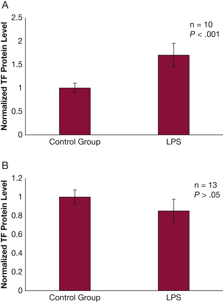
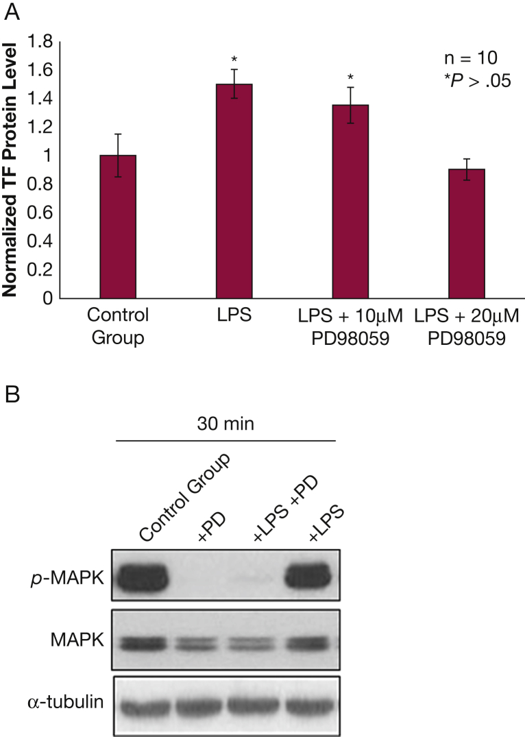
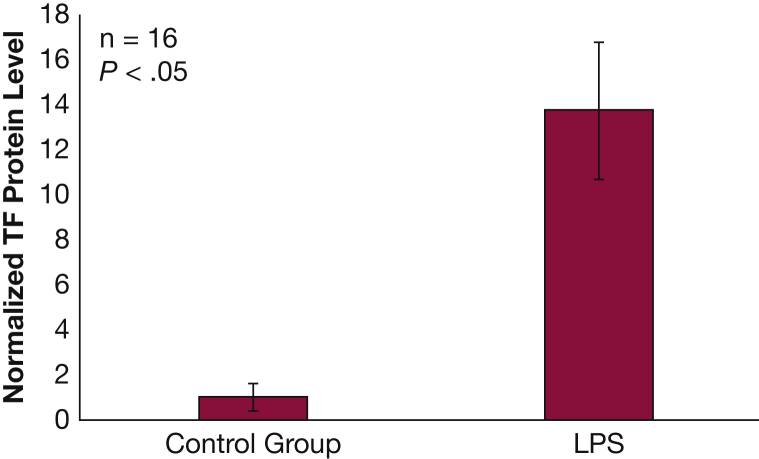
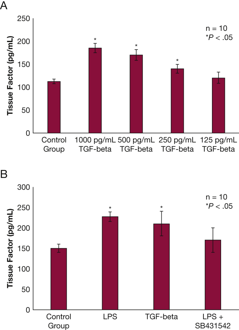
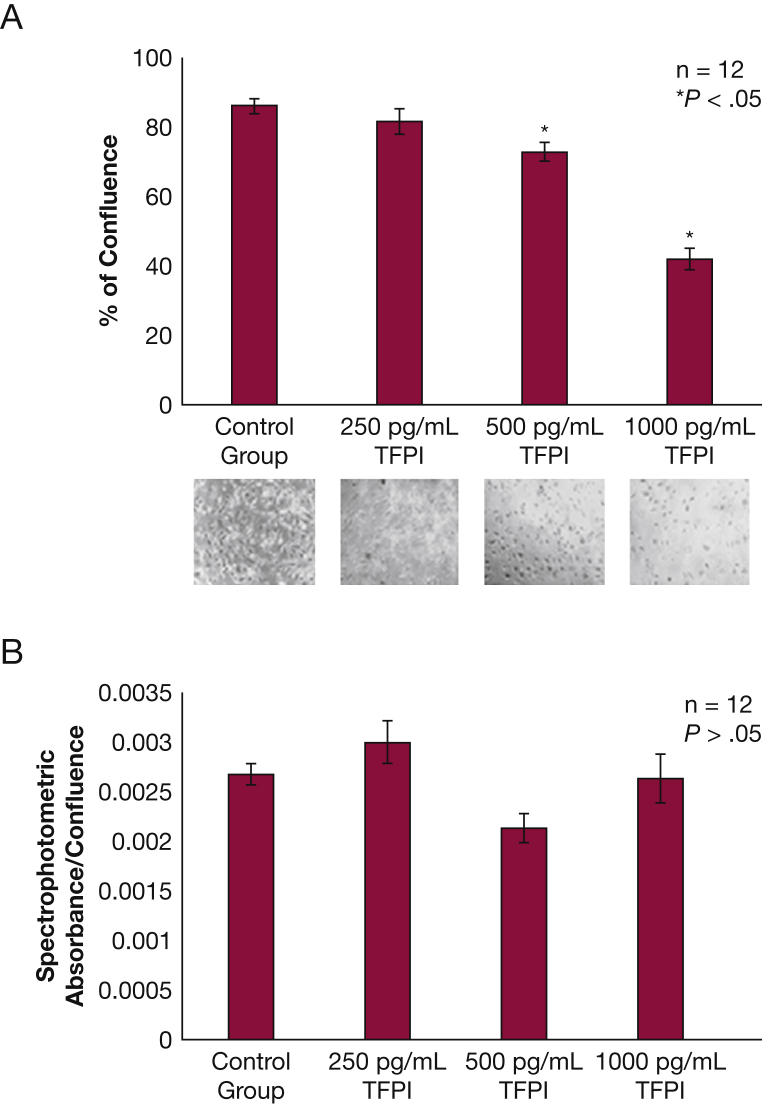
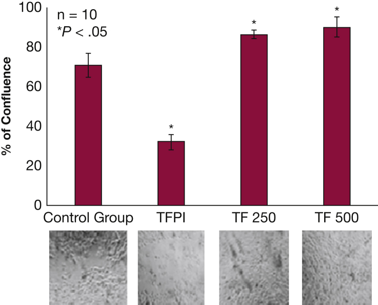
Results: TF protein levels increased in cell media after exposure to lipopolysaccharide (P < .01) but only in growing cells, and this action was blocked when exposed to an extracellular signal-regulated kinase inhibitor or a "small" worm phenotype and mothers against Decapentaplegic inhibitor. TF protein also increased in the presence of TGF-β (P < .05). Exposure to TF pathway inhibitor decreased the rate of cell growth by 60% (P < .05), and exposure to TF increased the rate of airway healing after injury by 19% (P < .05).

Conclusions: Growing airway epithelia release TF when exposed to lipopolysaccharide or TGF-β. TF reduces wound-healing time in airway epithelia and therefore may be important to airway recovery after injury.

Keywords: inflammation; remodeling; tissue factor; transforming growth factor β; wound healing.


Figures

Figure 1
Figure 1
Normal human bronchial epithelium releases tissue factor (TF) protein when exposed to LPS. A, Normal human bronchial epithelium exposed to 10 μg/mL of LPS for 24 h released > 50% more TF protein than those exposed to vehicle (P < .001). Three different cell lines were used to verify this observation, and data were normalized to reflect change vs control instead of reporting protein concentrations due to significant variation in relative concentrations between the cell lines. B, TF messenger RNA did not increase after exposure to 10 μg/mL of LPS for 3 h. Results normalized to reflect change vs control instead of reporting protein concentrations due to significant variation in relative concentrations between the cell lines. Results are reported as means with error bars indicating SEs. LPS = lipopolysaccharide.
Figure 2
Figure 2
–TF release from normal human bronchial epithelium (NHBE) following LPS exposure is mediated by mitogen-activated protein kinase. A, NHBE exposed to 10 μg/mL of LPS for 24 h release more TF than control (P < .05); NHBE exposed to 10 μg/mL of LPS for 24 h in the presence of 10 μM PD98059, a mitogen-activated protein kinase/extracellular signal-regulated kinase inhibitor, do not release more TF than control. B, PD98059 inhibits MAPK phosphorylation at baseline and after LPS exposure. See Figure 1 legend for expansion of other abbreviations.
Figure 3
Figure 3
NHBE transforming growth factor β protein levels increase following exposure to LPS. Levels of transforming growth factor β protein in cell lysate increased in NHBE after 24 h exposure to 10 μg/mL of LPS (P < .05). Results are reported as means with error bars indicating SEs. See Figure 1, Figure 2 legends for expansion of abbreviations.
Figure 4
Figure 4
TF release from NHBE following LPS exposure is mediated by TGF-β and “small” worm phenotype and mothers against Decapentaplegic inhibitor signaling. A, NHBE exposed to TGF-β for 24 h release TF in a dose-dependent manner (P < .05). B, When NHBE were exposed to LPS 10 μg/mL of or TGF-β 1 ng/mL for 24 h, they released more TF compared with control (P < .05). When cells were exposed to LPS 10 μg/mL + SB431542 (a “small” worm phenotype and mothers against Decapentaplegic inhibitor ) 20 μM, this inhibited TF release (P < .05). All results are reported as means with error bars indicating SEs. TGF-β = transforming growth factor β. See Figure 2, Figure 4 legends for expansion of other abbreviations.
Figure 5
Figure 5
NHBE exposed to TFPI do not reach confluence in culture. A, NHBE exposed to 1 ng/mL or 500 pg/mL of TFPI grew significantly slower under submerged conditions than those exposed to 250 pg/mL of TFPI and the control group (P < .05). Images of each exposure group are below their respective column in the bar graph. Results are reported as means with error bars indicating SEs. B, Lactate dehydrogenase levels were not significantly different between the control or treatment groups (P > .05). TFPI = tissue factor pathway inhibitor. See Figure 2 legend for expansion of other abbreviations.
Figure 6
Figure 6
TF decreases healing time in differentiated NHBE. NHBE differentiated as pseudostratified ciliated columnar epithelium in air-liquid interface conditions were scratched along the midline. Those exposed to 1 ng/mL of TFPI regrew more slowly than the control group (P < .05); those exposed to 250 or 500 pg/mL of TF regrew more rapidly than the control group (P < .05). Images of each exposure group are below their respective column in the bar graph. Results are reported as means with error bars indicating SEs. See Figure 1, Figure 2, Figure 5 legends for expansion of other abbreviations.

References

    1. Eilertsen K.E., Osterud B. Tissue factor: (patho)physiology and cellular biology. Blood Coagul Fibrinolysis. 2004;15(7):521–538. - PubMed
    1. Witkowski M., Landmesser U., Ranch U. Tissue factor as a link between inflammation and coagulation. Trends Cardiovasc Med. 2016;26(4):297–303. - PubMed
    1. Aberg M., Eriksson O., Siegbahn A. Tissue factor noncoagulant signaling: mechanisms and implications for cell migration and apoptosis. Semin Thromb Hemost. 2015;41(7):691–699. - PubMed
    1. Osterud B., Bjorklid E. Sources of tissue factor. Semin Thromb Hemost. 2006;32(1):11–23. - PubMed
    1. Mackman N. Role of tissue factor in hemostasis and thrombosis. Blood Cells Mol Dis. 2006;36(2):104–107. - PubMed

Publication types

MeSH terms