. 2018 Oct 22;9(10):514.
doi: 10.3390/genes9100514.

Genetic Polymorphisms and In Silico Mutagenesis Analyses of CYP2C9, CYP2D6, and CYPOR Genes in the Pakistani Population

Affiliations

Genetic Polymorphisms and In Silico Mutagenesis Analyses of CYP2C9, CYP2D6, and CYPOR Genes in the Pakistani Population

Shabbir Ahmed et al. Genes (Basel). .

Abstract

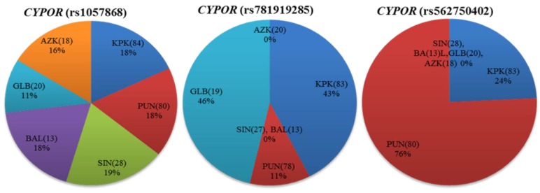
Diverse distributions of pharmacogenetically relevant variants of highly polymorphic CYP2C9, CYP2D6 and CYPOR genes are responsible for some varied drug responses observed across human populations. There is limited data available regarding the pharmacogenetic polymorphisms and frequency distributions of major allele variants in the Pakistani population. The present in silico mutagenesis study conducted on genotype pharmacogenetic variants and comparative analysis with a global population aims to extend the currently limited pharmacogenetic available evidence for the indigenous Pakistani population. Extracted genomic DNA from 244 healthy individuals' venous blood samples were amplified for distinct variant loci in the CYP2C9, CYP2D6 and CYPOR genes. Two-way sequencing results were compared with standard PubMed data and sequence variant loci confirmed by Chromas. This study revealed significant variations in CYP2C9 (rs1799853, rs1057910 and rs72558189), CYP2D6 (rs16947 and rs1135840), and CYPOR (rs1057868, rs781919285 and rs562750402) variants in intraethnic and interethnic frequency distributions. In silico mutagenesis and three-dimensional protein structural alignment analysis approaches clearly exposed the possible varied impact of rare CYPOR (rs781919285 and rs562750402) single nucleotide polymorphisms (SNPs) and confirmed that the influences of CYP2C9 and CYP2D6 variants are consistent with what was found in earlier studies. This investigation highlighted the need to study pharmacogenetic relevance loci and documentation since evidence could be utilized to elucidate genetic backgrounds of drug metabolism, and provide a basis for future pharmacogenomic studies and adequate dose adjustments in Pakistani and global populations.

Keywords: Pakistani population; alleles frequencies; drug-metabolizing enzymes; in silico mutagenesis; pharmacogenomics; polymorphism.


Conflict of interest statement

The authors declare no conflicts of interest.

Figures

Figure 1
Figure 1
Occurrence of cytochrome P450 2C9 (CYP2C9), cytochrome P450 2D6 (CYP2D6) and cytochrome P450 oxidoreductase (CYPOR) missense variants in the Pakistani cohort vs. eleven world populations represented in the 1000 Genomes Project. The p-value of the χ2 test in CYP2C9, CYP2D6 and CYPOR missense variants among the Pakistani cohort vs. global populations were calculated using the chi-square test and values were considered significant at the p < 0.05 level. Statistically significant variations are represented by a blue bar (af). Global populations are arranged according to the least significant to most significant negative log(p-value). Populations included were African ancestry in Southwest USA (ASW); Luhya in Webuye, Kenya (LWK); Yoruba in Ibadan, Nigeria (YRI); Mexican ancestry in Los Angeles, CA, USA (MEX); Han Chinese in Beijing, China (CHB); Southern Han Chinese, China (CHS); Japanese in Tokyo, Japan (JPT); Utah, USA residents with Northern and Western European ancestry from the Centre d’Etude du Polymorphisme Humain (CEPH) collection (CEU); Toscani in Italy (TSI); Gujarati Indians in Houston, Texas, USA (GIH); Sri Lankan Tamal in UK (STU). Numbers in parentheses indicate the group size.
Figure 2
Figure 2
CYP2C9*2, CYP2C9*3, and CYP2C9*14 variant allele distributions in indigenous Pakistani populations. The distribution of the percentage allele frequency of pharmacogenetic CYP2C9*2, CYP2C9*3 and CYP2C9*14 variants in Pakistani local populations. Regional populations are represented by colored columns in decreasing variant allele percentage frequency order. The CYP2C9*2, CYP2C9*3, and CYP2C9*14 variants are associated with reduced enzyme metabolic activity in comparison to the CYP2C9*1 wild-type. The CYP2C9*2 variant was found most frequently in the AZK region while CYP2C9*3 and CYP2C9*14 allele variants were in relatively higher frequencies in individuals from KPK. However, these variants were least frequent in GLB territory, as well as CYP2C9*2. KPK: Khyber Pakhtunkhwa; PUN: Punjab; SIN: Sindh; BAL: Balochistan; GLB: Gilgit-Baltistan; and AZK: Azad Kashmir. Numbers in parentheses indicate the group size.
Figure 3
Figure 3
The allele frequency distribution of CYP2C9*2, CYP2C9*3, and CYP2C9*14 variants in indigenous Pakistani and global populations. Percentage allele frequency distributions of CYP2C9*2 (rs1799853), CYP2C9*3 (rs1057910) and CYP2C9*14 (rs72558189) pharmacogenetic variants in the Pakistani population and global populations from the 1000 Genome Project depicted in bar chart plots. Frequencies in the Pakistani population are shown in yellow, while others are represented by blue colored columns in the order of decreasing variant allele percentage frequency. The CYP2C9*2, CYP2C9*3, and CYP2C9*14 three CYP2C9 missense variants associated with reduced enzyme activities, were found to be almost the least frequent, intermediately frequent, and highly frequent respectively, in indigenous Pakistani individuals compared with global populations. PAK: Pakistani, Pakistan; ASW: African ancestry in Southwest USA; LWK: Luhya in Webuye, Kenya; YRI: Yoruba in Ibadan, Nigeria; MXL: Mexican ancestry in Los Angeles, California, USA; CHB: Han Chinese in Beijing, China; CHS: Southern Han Chinese, China; JPT: Japanese in Tokyo, Japan; CEU: Utah, USA residents with Northern and Western European ancestry from the CEPH collection; TSI: Toscani in Italy; GIH: Gujarati Indians in Houston, Texas, USA; STU: Sri Lankan Tamal in UK. Numbers in parentheses indicate the group size.
Figure 4
Figure 4
Distribution of the cytochrome P450 oxidoreductase (CYPOR) missense variants among Pakistani indigenous populations. Percentage allele frequency distributions of CYPOR (rs1057868, rs781919285 and rs562750402) are shown as pie charts for different regions of Pakistan. Numbers in parentheses indicate the group size.
Figure 5
Figure 5
The structural alignment of human CYP2C9 (PDB: 1OG2) to the substrate free mutant CYP2C9 protein obtained by in silico mutagenesis. Hydrogen bonds are represented as a blue dotted line and the water molecule as hot pink spheres. The heme domain is shown in blue, while arrows indicate the deviation sites among the structures of normal (green) and mutated (red) proteins found in the Pakistani population. The wheat colored a, c, and e, and cyan b, d, and f show the 3D representations of normal and altered hydrogen bond interactions corresponding to amino acid changes, respectively. (a) a close view of arginine amino acid at the position of 144 intermolecular interactions. The residue has an intensive hydrogen bond network with the R139, S140, I141, E147, E148, S180 and Q261 backbone polypeptide amino acids and a tightly bounded water molecule; (b) visualization of hydrogen bond networking perturbation in the CYP2C9 crystal structure, produced by in silico mutagenesis of Arg144 with a relatively small 144Cys residue. This in silico mutagenesis resulted in the breaking of three intermolecular hydrogen interactions, with the adjacent arginine 139, glutamine 261, and serine 180 leading to altered CYP2C9 protein interaction with CYPOR [9]; (c) intramolecular hydrogen bonding of wild-type Ile359 located in the substrate recognition site. It is important in substrate specificity and affinity determination [75]; (d) in silico produced missense mutation visualization in the CYP2C9 crystal structure by the substitution of Ile359 with Lue 359; (e) Arg125 lies near the heme domain, on the solvent-exposed protein surface, and interacts with NADPH-P450 reductase in NADPH-dependent metabolisms [75]; (f) a partial view of the missense mutation resulting in the replacement of Arg125 with 125His. This leads to varied interactions necessary for NADPH-dependent CYP2C9 metabolic activity [76].
Figure 6
Figure 6
Structural comparison of human CYP2D6 (PDB: 2F9Q) to the mutant CYP2D6 protein depicting normal and missense mutation sites reported in the Pakistani population. Wild-type CYP2D6 is represented in wheat, while mutant-type is represented in cyan. Positions of normal (green) and mutant (red) amino acid residues are indicated by black arrow heads. (a) the hydrogen interactions of wild-type Arg296 residue to peptide backbone Asp292 and Ala300 residues; (b) X-ray structure partial view of missense SNP produced by in silico mutagenesis through the substitution of positively charged polar Arg296 with the relatively smaller nonpolar Cys296 residue; (c,d) highlight hydrogen bond interactions of wild-type Ser486 and mutant Thr486 amino acids with the adjacent Val480 residue in peptide backbone, represented in wheat and cyan, respectively.
Figure 7
Figure 7
A secondary structural feature-based (helices in red, sheets in yellow and loops in green) schematic illustration of the human cytochrome P450 oxidoreductase (CYPOR). The co-factors FAD (magenta), NADP(H) (blue), and FMN (orange) are shown as ball and stick models. Missense mutations identified in the Pakistani population are shown as spheres.
Figure 8
Figure 8
Three-dimensional structural alignment of wild-type human CYPOR (PDB: 3QE2) and mutant protein produced by in silico mutagenesis, showing missense mutations corresponding to the Pakistani population (wheat color) and wild-type (cyan color) peptide backbones. Cartoon structures of human CYPOR are depicted, as well as cofactors FMN (blue-gray) and FAD (yellow) and coenzyme NADP(H) (pink) in stick configuration. The interaction analysis of the wild-type and mutant residues strongly suggest that the two structures of human CYPOR are almost identical, except for the sites of missense SNPs. Partial views of crystal structures show a hydrogen bonding network involving water molecules (hot pink) and peptide backbones (blue dotted lines). (a) the wild-type Thr91 residue shows three hydrogen interactions each with the adjacent flavin mononucleotide (FMN) cofactor and water molecules; (b) the substitution of polar Thr91 with the nonpolar Met91 residue resulted in the loss of three hydrogen interactions, one with FMN and two with water molecules that were involved in its stabilization; (c) wild-type Glu95 directly interact with Asn99, Lys360 residues, and water molecules via hydrogen bonding; (d) a change in the hydrogen bonding network is observed due to the replacement of the negatively charged Glu95 with the nonpolar Gly95 residue; (e) hydrogen interaction between wild-type Ala503 and adjacent nonpolar Val289 residue; (f) apparently, the replacement of wild type Ala503 residue with Val503 does not affect the bonding interactions with the peptide backbone.

References

    1. Bernard S., Neville K.A., Nguyen A.T., Flockhart D.A. Interethnic differences in genetic polymorphisms of CYP2D6 in the U.S. Population: Clinical implications. Oncologist. 2006;11:126–135. doi: 10.1634/theoncologist.11-2-126. - DOI - PubMed
    1. Ma M.K., Woo M.H., Mcleod H.L. Genetic basis of drug metabolism. Am. J. Health-Syst. Pharm. 2002;59:2061–2069. - PubMed
    1. Zhang H.F., Li Z.H., Liu J.Y., Liu T.T., Wang P., Fang Y., Zhou J., Cui M.Z., Gao N., Tian X., et al. Correlation of cytochrome P450 oxidoreductase expression with the expression of 10 isoforms of cytochrome P450 in human liver. Drug Metab. Dispos. 2016;44:1193–1200. doi: 10.1124/dmd.116.069849. - DOI - PMC - PubMed
    1. Gardiner S.J., Begg E.J. Pharmacogenetics, drug-metabolizing enzymes, and clinical practice. Pharmacol. Rev. 2006;58:521–590. doi: 10.1124/pr.58.3.6. - DOI - PubMed
    1. Lee C.R., Goldstein J.A., Pieper J.A. Cytochrome P450 2C9 polymorphisms: A comprehensive review of the in-vitro and human data. Pharmacogenetics. 2002;12:251–263. doi: 10.1097/00008571-200204000-00010. - DOI - PubMed

LinkOut - more resources