Review
. 2018 Oct 23;8(4):120.
doi: 10.3390/biom8040120.

Modeling Membrane Curvature Generation due to Membrane⁻Protein Interactions

Affiliations
Review

Modeling Membrane Curvature Generation due to Membrane⁻Protein Interactions

Haleh Alimohamadi et al. Biomolecules. .

Abstract

To alter and adjust the shape of the plasma membrane, cells harness various mechanisms of curvature generation. Many of these curvature generation mechanisms rely on the interactions between peripheral membrane proteins, integral membrane proteins, and lipids in the bilayer membrane. Mathematical and computational modeling of membrane curvature generation has provided great insights into the physics underlying these processes. However, one of the challenges in modeling these processes is identifying the suitable constitutive relationships that describe the membrane free energy including protein distribution and curvature generation capability. Here, we review some of the commonly used continuum elastic membrane models that have been developed for this purpose and discuss their applications. Finally, we address some fundamental challenges that future theoretical methods need to overcome to push the boundaries of current model applications.

Keywords: Helfrich energy; area difference elastic model; deviatoric curvature; hydrophobic mismatch; plasma membrane; protein crowding; spontaneous curvature.


Conflict of interest statement

The authors declare no conflict of interest.

Figures
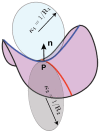
Figure A1
Figure A1
Principal curvatures of a surface.
Figure A2
Figure A2
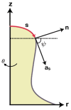
Axisymmetric coordinates with z as the axis of rotation.
Figure 1
Figure 1
Membrane curvature generation in cells and associated modeling results: (A) Membrane budding in endocytosis. Reprinted with permission from references [66,67]. Copyright 2017 PNAS and Copyright 2018 PCCP; (B) Formation and stabilization of tubular membrane structures in the Golgi. Reprinted with permission from references [68,69]. Copyright 2013 PloS one and Copyright 2017 ACS Nano; (C) Change in the topology of nuclear envelopes. Reprinted with permission from reference [70]. Copyright 2016 PNAS; (D) Membrane invagination in caveolae. Reprinted with permission from references [71,72]. Copyright 2013 Soft matter and Copyright 2011 J. Phys. Chem. B.; (E) Actin force driven filopodia protrusion. Reprinted with permission from references [73,74]. Copyright 2015 PNAS and Copyright 2016 PLoS Comput Biol.; and (F) Mitochondrial fission. Reprinted with permission from references [75,76]. Copyright 2017 Front Physiol and Copyright 2007 J. Phys. Chem. B.
Figure 2
Figure 2
Schematic depiction of a cellular membrane highlighting its composition. There are two layers of amphipathic lipid molecules that self-assemble to form the bilayer. In each layer, the hydrophilic head groups form the outer surface and the hydrophobic tails face toward each other in the interior region. The distribution and organization of lipids and different proteins can vary from cell to cell. The cell membrane is composed of many different molecules including peripheral proteins, integral proteins, and carbohydrate molecules.
Figure 3
Figure 3
Cartoon models of the mechanisms of membrane curvature generation due to protein (shown in red) interactions in different continuum elastic models. (A) Local protein interactions with membrane produce a spontaneous curvature field. s is the arc length parameterization along the membrane and C is the induced spontaneous curvature. (B) The asymmetric insertion of conical proteins on one side of the membrane results in the expansion of the upper leaflet and compression of the lower leaflet. (C) Asymmetric insertion of proteins into the lipid bilayer induces both local spontaneous curvature and surface stresses due to membrane leaflets expansion/compression. (D) Rotationally non-symmetric proteins generate anisotropic curvature. (E) Aggregated proteins on the membrane surface create a spontaneous curvature field and also have entropic interactions with the membrane. Here, ϕ represents the relative density of the accumulated proteins. (F) The induced pressure (p) by crowding proteins drives membrane bending. a is the surface area occupied by one protein.
Figure 4
Figure 4
Mattress model representation of the hydrophobic mismatch phenomena [208]. Both protein and lipid bilayer are modeled as one-dimensional springs with constants Ap and Al, respectively. dl0 and dp0 are the initial lengths, and dl and dp are the final lengths of the lipid bilayer and the protein after deformation, respectively. The gray area corresponds to the hydrophilic region with the strength of Blp, and the yellow region indicates the adhesive region with adhesive interactions strength of Clp.
Figure 5
Figure 5
Perspective for the future of theoretical models for membrane curvature generating mechanisms. (A) Various mechanisms are involved in trafficking including amphipathic helix insertion into the bilayer, protein scaffolding, cargo-receptor crowding, forces from actin polymerization, and lipid phase separation [236,237]. (B) The coupling between membrane shape, membrane curvature, and membrane proteins distribution. The convex proteins (indicated with red cones) aggregate and flow toward the hill where the membrane curvature is negative (assuming the normal vector to the surface is outward). On the other hand, the concave proteins (represented by blue cones) accumulate and move toward the valley where the membrane curvature is large and positive [176]. (C) The coupling between the formation of a filopodial protrusion and the intracellular signaling inside the cell [238]. The ligand attachment to the G-protein-coupled receptor (GPCR) activates isotype β of the phospholipase C (PLCβ) which is a class of membrane-associated enzymes. PLCβ stimulates the phosphatidylinositol 4,5–bisphosphate (PIP2) which is a phospholipid component of the cell membrane and regulates the membrane tension. The hydrolysis of PIP2 produces the messenger molecule inositol trisphosphate (IP3). Binding IP3 molecules to the ER releases the calcium (Ca2+) that stored in the ER to the cytoplasm. The Ca2+ is a key intracellular molecule that controls the actin polymerization at the leading edge of the membrane protrusion.

References

    1. Mukherjee S., Maxfield F.R. Role of membrane organization and membrane domains in endocytic lipid trafficking. Traffic. 2000;1:203–211. doi: 10.1034/j.1600-0854.2000.010302.x. - DOI - PubMed
    1. Gruenberg J. The endocytic pathway: A mosaic of domains. Nat. Rev. Mol. Cell Biol. 2001;2:721. doi: 10.1038/35096054. - DOI - PubMed
    1. Westermann B. Mitochondrial fusion and fission in cell life and death. Nat. Rev. Mol. Cell Biol. 2010;11:872. doi: 10.1038/nrm3013. - DOI - PubMed
    1. Van der Bliek A.M., Shen Q., Kawajiri S. Mechanisms of mitochondrial fission and fusion. Cold Spring Harb. Perspect. Biol. 2013;5:a011072. doi: 10.1101/cshperspect.a011072. - DOI - PMC - PubMed
    1. Grafmuller A., Shillcock J., Lipowsky R. The fusion of membranes and vesicles: Pathway and energy barriers from dissipative particle dynamics. Biophys. J. 2009;96:2658–2675. doi: 10.1016/j.bpj.2008.11.073. - DOI - PMC - PubMed

Publication types

MeSH terms

Substances