. 2018 Oct 23;10(4):199.
doi: 10.3390/pharmaceutics10040199.

Sterically Stabilized RIPL Peptide-Conjugated Nanostructured Lipid Carriers: Characterization, Cellular Uptake, Cytotoxicity, and Biodistribution

Affiliations

Sterically Stabilized RIPL Peptide-Conjugated Nanostructured Lipid Carriers: Characterization, Cellular Uptake, Cytotoxicity, and Biodistribution

Chang Hyun Kim et al. Pharmaceutics. .

Abstract

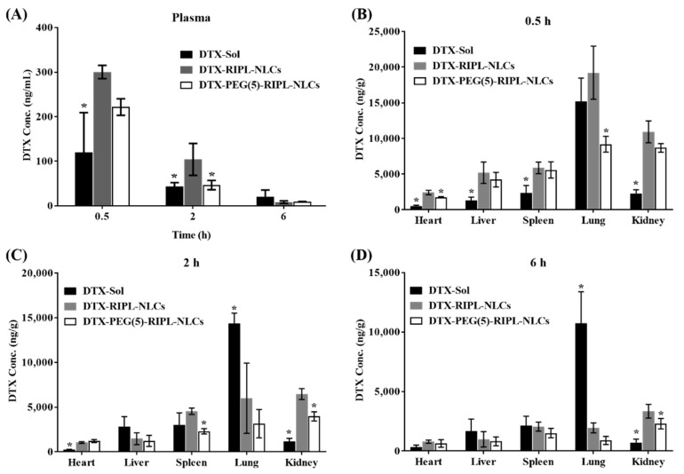
As a platform for hepsin-specific drug delivery, we previously prepared IPLVVPLRRRRRRRRC peptide (RIPL)-conjugated nanostructured lipid carriers (RIPL-NLCs) composed of Labrafil® M 1944 CS (liquid oil) and Precirol® ATO 5 (solid lipid). In this study, to prevent the recognition by the mononuclear phagocyte system, polyethylene glycol (PEG)-modified RIPL-NLCs (PEG-RIPL-NLCs) were prepared using PEG3000 at different grafting ratios (1, 5, and 10 mole %). All prepared NLCs showed a homogeneous dispersion (130⁻280 nm), with zeta potentials varying from -18 to 10 mV. Docetaxel (DTX) was successfully encapsulated in NLCs: encapsulation efficiency (93⁻95%); drug-loading capacity (102⁻109 µg/mg). PEG-RIPL-NLCs with a grafting ratio of 5% PEG or higher showed significantly reduced protein adsorption and macrophage phagocytosis. The uptake of PEG(5%)-RIPL-NLCs by cancer cell lines was somewhat lower than that of RIPL-NLCs because of the PEG-induced steric hindrance; however, the uptake level of PEG-RIPL-NLCs was still greater than that of plain NLCs. In vivo biodistribution was evaluated after tail vein injection of NLCs to normal mice. Compared to RIPL-NLCs, PEG(5%)-RIPL-NLCs showed lower accumulation in the liver, spleen, and lung. In conclusion, we found that PEG(5%)-RIPL-NLCs could be a promising nanocarrier for selective drug targeting with a high payload of poorly water-soluble drugs.

Keywords: RIPL peptide; biodistribution; cellular uptake; cytotoxicity; nanostructured lipid carrier; steric stabilization.


Conflict of interest statement

The authors declare that they have no conflicts of interests.

Figures

Figure 1
Figure 1
Conformational comparison of different nanostructured lipid carriers (NLCs).
Figure 2
Figure 2
Characterization of various NLCs. (A) TEM images of docetaxel (DTX)-loaded NLCs. Scale bar = 200 nm. (B) Conjugation efficiencies of the RIPL peptide. Data represent the mean ± SD (n = 3). (C) In vitro release profiles of DTX from various formulations. Data are the mean ± SD (n = 3).
Figure 3
Figure 3
Bovine serum albumin (BSA) adsorption on various NLCs as a function of time. * p < 0.05 versus RIPL peptide-conjugated NLCs (RIPL-NLCs); # p < 0.05 versus PEG (1 mole %)-grafted RIPL-NLCs (PEG(1)-RIPL-NLCs).
Figure 4
Figure 4
Phagocytic uptake of various 1,1′-dioctadecyl-3,3,3′,3′-tetramethylindocarbocyanine perchlorate (DiI)-loaded NLCs by RAW 264.7 macrophage cells. (A) Confocal laser scanning microscopy (CLSM) images after 2 h incubation. The nucleus is stained with 4′,6-diamidino-2-phenylindole (DAPI) (blue), and DiI is distributed in the cells (red). Scale bar = 20 μm. (B) Flow cytometry histogram of phagocytosed DiI-loaded NLCs after 2 h incubation. (C) CLSM images acquired after treatment with RIPL-NLCs and PEG(5)-RIPL-NLCs for different times. Scale bar = 20 μm. (D) MFI values calculated after treatment with RIPL-NLCs and PEG (5 mole %)-grafted RIPL-NLCs (PEG(5)-RIPL-NLCs). Data are the mean ± SD (n = 3). * p < 0.05.
Figure 5
Figure 5
Cellular uptake of DiI-loaded NLCs by the SKOV3, MCF7, and LNCaP cell lines. (A) Flow cytometry histogram of plain NLCs (pNLCs), RIPL-NLCs, and PEG(5)-RIPL-NLCs uptake. (B) The relative MFI values of treatments: RIPL-NLCs versus pNLCs and PEG(5)-RIPL-NLCs versus pNLCs. Data are the mean ± SD (n = 3). (C) CLSM images of SKOV3 cells incubated with DiI-loaded NLCs. Scale bar = 20 μm.
Figure 6
Figure 6
Competitive inhibition study of cellular uptake of RIPL-NLCs and PEG(5)-RIPL-NLCs by SKOV3 cells in the presence of endocytosis inhibitors and at a low temperature. Data are the mean ± SD (n = 3). * p < 0.05 versus control; # p < 0.05 between RIPL-NLCs and PEG(5)-RIPL-NLCs.
Figure 7
Figure 7
Cytotoxicity of the empty and DTX-loaded formulations for SKOV3, MCF7, and LNCaP cells. (A) Cell viability plotted against the log DTX-equivalent concentration. DTX-Sol (○,●); DTX-pNLCs (□,∎); DTX-RIPL-NLCs (△,▲); DTX-PEG(5)-RIPL-NLCs (◊,♦). Opened for empty formulations and filled for DTX-loaded formulations; (B) IC50 values of DTX-loaded formulations. Data are the mean ± SD (n = 3). * p < 0.05 versus DTX-pNLCs.
Figure 8
Figure 8
Hemocompatibility of the empty and DTX-loaded formulations with red blood cells (RBCs). (A) Photographs of tubes after centrifugation. (B) Percentage of hemolysis within 1 h. A saline solution was used as the negative control, and Triton X-100 (1%, v/v) was added to the positive control. Data are the mean ± SD (n = 3).
Figure 9
Figure 9
Biodistribution of DTX-loaded formulations in Institute of Cancer Research (ICR) mice after tail vein injection at a dose of 5 mg DTX-equivalent/kg (∎: DTX-Sol; : DTX-RIPL-NLCs; □: DTX-PEG(5)-RIPL-NLCs). Concentrations of DTX in the plasma (A) and representative organs are presented at different time points (0.5 h (B), 2 h (C), and 6 h (D)). Data are the mean ± SD (n = 5). * p < 0.05 versus DTX-RIPL-NLCs.

Similar articles

Cited by

References

    1. Iqbal M.A., Asif M.D., Sahni J.K., Baboota S., Dang S., Ali J. Nanostructured lipid carriers system: Recent advances in drug delivery. J. Drug Target. 2012;20:813–830. doi: 10.3109/1061186X.2012.716845. - DOI - PubMed
    1. Zhang X., Pan W., Gan L., Zhu C., Gan Y., Nie S. Preparation of a dispersible PEGylate nanostructured lipid carriers (NLC) loaded with 10-hydroxycamptothecin by spray-drying. Chem. Pharm. Bull. 2008;56:1645–1650. doi: 10.1248/cpb.56.1645. - DOI - PubMed
    1. Yang Y., Yang Y., Xie X., Cai X., Mei X. Preparation and characterization of photo-responsive cell-penetrating peptide-mediated nanostructured lipid carrier. J. Drug Target. 2014;22:891–900. doi: 10.3109/1061186X.2014.940589. - DOI - PubMed
    1. Liu D., Liu F., Liu Z., Wang L., Zhang N. Tumor specific delivery and therapy by double-targeted nanostructured lipid carriers with anti-VEGFR-2 antibody. Mol. Pharm. 2011;8:2291–2301. doi: 10.1021/mp200402e. - DOI - PubMed
    1. Kang M.H., Park M.J., Yoo H.J., Hyuk K.Y., Lee S.G., Kimm S.R., Yeom D.W., Kang J.K., Choi Y.W. RIPL peptide (IPLVVPLRRRRRRRRC)-conjugated liposomes for enhanced intracellular drug delivery to hepsin-expressing cancer cells. Eur. J. Pharm. Biopharm. 2014;87:489–499. doi: 10.1016/j.ejpb.2014.03.016. - DOI - PubMed