. 2018:82:141-195.
doi: 10.1016/bs.ctm.2018.09.003. Epub 2018 Oct 15.

Cortical Actin Dynamics in Endothelial Permeability

Affiliations

Cortical Actin Dynamics in Endothelial Permeability

Patrick Belvitch et al. Curr Top Membr. 2018.

Abstract

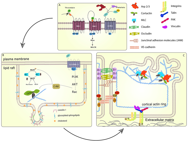
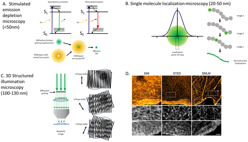
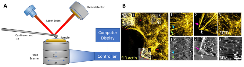
The pulmonary endothelial cell forms a critical semi-permeable barrier between the vascular and interstitial space. As part of the blood-gas barrier in the lung, the endothelium plays a key role in normal physiologic function and pathologic disease. Changes in endothelial cell shape, defined by its plasma membrane, determine barrier integrity. A number of key cytoskeletal regulatory and effector proteins including non-muscle myosin light chain kinase, cortactin, and Arp 2/3 mediate actin rearrangements to form cortical and membrane associated structures in response to barrier enhancing stimuli. These actin formations support and interact with junctional complexes and exert forces to protrude the lipid membrane to and close gaps between individual cells. The current knowledge of these cytoskeletal processes and regulatory proteins are the subject of this review. In addition, we explore novel advancements in cellular imaging that are poised to shed light on the complex nature of pulmonary endothelial permeability.

Keywords: ARDS; Arp 2/3; Atomic force microscopy; Cortactin; Cortical actin; Endothelial permeability; Intravital microscopy; Lamellipodia; Lung injury; Non-muscle myosin light chain kinase; Super-resolution microscopy.


Figures

Figure 1.
Figure 1.. Barrier regulation by cytoskeletal and membrane structures.
Pulmonary endothelial barrier integrity is the result of coordinated cell processes involving receptors, signaling molecules, junctional complexes and protein-regulated cytoskeletal rearrangements leading to changes in membrane dynamics. (A) Activation of specific transmembrane receptors are critical to initiate barrier disruptive (PAR-1) or protective (S1PR1) signaling events. (B) Ligation of the S1PR1 receptor recruits signaling molecules and cytoskeletal effector proteins to lipid rafts. Phosphorylation of cortactin and myosin light chain kinase results in rapid cytoskeletal changes. (C) The formation of a cortical actin ring provides structural stability and anchoring for multiple membrane-bound junctional complexes which become activated, strengthening connections to neighboring cells and the extra cellular matrix. Branched actin polymerization, regulated by Arp 2/3 and cortactin, generates protrusive force on the plasma membrane, forming sheet-like projections or lamellipodia which close gaps between individual cells and further enhance membrane junctions.
Figure 2.
Figure 2.. Super-resolution microscopy techniques.
The fluorescence and optical principles for three super-resolution microscopy techniques are shown with demonstrated lateral resolution noted in parentheses. (A) Stimulated emission depletion microscopy (STED): Effectively reduces the volume of the point spread function (PSF) of an objective lens, which is the three-dimensional diffraction volume of emitted light transmitted by an objective lens from an infinitesimally small point source of light in the specimen. A red-shifted laser with hollow beam (depletion laser) quenches fluorophores on the periphery of the PSF, leaving a small central subdiffraction volume of excited fluorophores that emit photons. Adapted from Blom and Widegren, 2017. (B) Single molecule localization microscopy (SMLM): Encompasses several related techniques that obtain many thousands of images of a given sample in which only a very small number of fluorophores are fluorescing at a given time. Precise localization of the geometric centers of individual fluorophores within the thousands of collected images can then be used to reconstruct a super-resolution image. (C) Structured illumination microscopy (SIM): A moveable diffraction grating is placed in the illumination aperture of the excitation beam path of a laser-illuminated wide-field microscopy set-up to generate a sinusoidal pattern of light with resulting Moiré fringes. The high frequency illumination stripes and high frequency object organization create even higher spatial frequencies below the diffraction limit. Multiple raw images must be collected at different diffraction orientations to reconstruct the final super-resolved image. (D) Actin cytoskeleton of COS-7 cells labeled with phalloidin-AlexaFluor 488 using SIM and STED and phalloidin-AlexaFluor647 using SMLM. Boxed areas note higher resolution areas of the lamella and lamellipodium in each optical section. Reprinted with permission from Wegel et al., 2016.
Figure 3.
Figure 3.. Combined super-resolution and atomic force microscopy.
(A) Components of a typical atomic force microscope (AFM) are depicted. AFM employs an exquisitely sensitive cantilever upon which a laser beam is reflected and a photodetector senses laser beam deflections to map the cell surface at atomic resolution of 0.1 nm in both lateral and axial dimensions. Adapted from Shan and Wang, 2015. (B) Representative image of a live murine astrocyte obtained by sequential, correlated STED-AFM microscopy. Super-resolution STED images (top inset) reveal polarized F-actin in the lamella and near the leading edge. Actin labelled with SiR-actin can be seen with corresponding thick filaments in AFM images (bottom panel) associated with focal adhesions (arrows). Reprinted with permission from Curry, Ghezali et al. 2017.

Similar articles

Cited by

References

    1. Abbasi T and Garcia JG (2013). “Sphingolipids in lung endothelial biology and regulation of vascular integrity.” Handb Exp Pharmacol(216): 201–226. - PMC - PubMed
    1. Abu Taha A and Schnittler HJ (2014). “Dynamics between actin and the VE-cadherin/catenin complex: novel aspects of the ARP2/3 complex in regulation of endothelial junctions.” Cell Adh Migr 8(2): 125–135. - PMC - PubMed
    1. Abu Taha A, Taha M, Seebach J and Schnittler HJ (2014). “ARP2/3-mediated junction-associated lamellipodia control VE-cadherin-based cell junction dynamics and maintain monolayer integrity.” Mol Biol Cell 25(2): 245–256. - PMC - PubMed
    1. Adderley SP, Lawrence C, Madonia E, Olubadewo JO and Breslin JW (2015). “Histamine activates p38 MAP kinase and alters local lamellipodia dynamics, reducing endothelial barrier integrity and eliciting central movement of actin fibers.” Am J Physiol Cell Physiol 309(1): C51–59. - PMC - PubMed
    1. Adyshev DM, Dudek SM, Moldobaeva N, Kim KM, Ma SF, Kasa A, Garcia JG and Verin AD (2013). “Ezrin/radixin/moesin proteins differentially regulate endothelial hyperpermeability after thrombin.” Am J Physiol Lung Cell Mol Physiol 305(3): L240–255. - PMC - PubMed

Publication types