Review
. 2018 Oct 30;72(18):2198-2212.
doi: 10.1016/j.jacc.2018.08.2150.

Monocyte and Macrophage Dynamics in the Cardiovascular System: JACC Macrophage in CVD Series (Part 3)

Affiliations
Review

Monocyte and Macrophage Dynamics in the Cardiovascular System: JACC Macrophage in CVD Series (Part 3)

Zahi A Fayad et al. J Am Coll Cardiol. .

Abstract

It has long been recognized that the bone marrow is the primary site of origin for circulating monocytes that may later become macrophages in atherosclerotic lesions. However, only in recent times has the complex relationship among the bone marrow, monocytes/macrophages, and atherosclerotic plaques begun to be understood. Moreover, the systemic nature of these interactions, which also involves additional compartments such as extramedullary hematopoietic sites (i.e., spleen), is only just becoming apparent. In parallel, progressive advances in imaging and cell labeling techniques have opened new opportunities for in vivo imaging of monocyte/macrophage trafficking in atherosclerotic lesions and at the systemic level. In this Part 3 of a 4-part review series covering the macrophage in cardiovascular disease, the authors intersect systemic biology with advanced imaging techniques to explore monocyte and macrophage dynamics in the cardiovascular system, with an emphasis on how events at the systemic level might affect local atherosclerotic plaque biology.

Keywords: atherosclerosis; bone marrow; cardiovascular; imaging; macrophage.


Conflict of interest statement

Disclosures:

The authors declare no conflicts of interest.

Figures
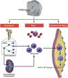
Central Illustration:
Central Illustration:. Systemic pathways and feedback loops regulating monocyte and macrophage trafficking in atherosclerosis, and the role of imaging.
The bone marrow is the primary site for the production of monocytes, which are generated from HSPCs. Once produced, monocytes are mobilized to the blood. HSPCs likewise mobilize to the blood according to circadian fluctuations. In the context of inflammation, HSPCs may give rise to monocytes in extramedullary sites such as the spleen. Bone marrow and spleen-derived monocytes circulate and infiltrate atherosclerotic lesions. Ly6chigh monocytes preferentially enter lesions where they differentiate to macrophages that can produce interleukin-1β (IL-1β), among other inflammatory products. Lesional macrophages can then proliferate and thus aggravate atherosclerosis. The various imaging approaches described in this review have shown enormous promise for understanding these monocyte/macrophage pathways at the systemic level.
Figure 1:
Figure 1:. Schematic depiction of contrast agents and tracers that can be used to map monocyte recruitment (R), and the accumulation (A), proliferation (P) and egress (E) of macrophages from the vessel wall, as well as metabolic activity in hematopoietic organs (bone marrow and spleen).
Radiolabeled nanobodies and peptides, iron oxides or gadolinium based agents targeted towards adhesion molecules (i.e. VCAM-1, P-selectin) are able to quantify monocyte vessel wall recruitment. Several radiotracers (18F-FDG, 68Ga-DOTATATE, 18F mannose, 18F-NaF) and MRI or CT contrast agents (iron oxides, gold nanoparticles) can be used to quantify macrophage plaque accumulation. Macrophage proliferation can be quantified with the thymidine analog 18F-FLT, while fluorescent microbeads have been reported as a marker of egress.
Figure 2:
Figure 2:. In vivo imaging of monocyte recruitment, and plaque macrophage accumulation, proliferation and egress.
Monocyte recruitment (upper left panel) can be probed using DCEMRI to quantify endothelial permeability (148,149), or adhesion molecule imaging with targeted iron oxides for MRI or antibodies and nanobodies for PET imaging (79,81). Macrophage plaque accumulation (top right panel) can be quantified using 18F-FDG PET (90,150), or iron oxide MRI (120). Macrophage proliferation (bottom left panel) in plaque or hematopoietic organs (bone marrow and spleen) can be instead measured 18F-FLT (136). While no in vivo methods are currently available to image macrophage plaque egress (lower right panel), fluorescent beads (arrows) have been previously used for this purpose in terminal experiments (139). All images were obtained with permission from the respective publishers associated with these citations.

References

    1. Nabel EG, Braunwald E. A tale of coronary artery disease and myocardial infarction. N Engl J Med 2012;366:54–63. - PubMed
    1. Bloom D, Cafiero E, Jané-Llopis E. The Global Economic Burden of Non-communicable Diseases Cambridge, MA: Harvard School of Public Health, 2011.
    1. Ridker PM, Everett BM, Thuren T et al. Antiinflammatory Therapy with Canakinumab for Atherosclerotic Disease. N Engl J Med 2017;377:1119–1131. - PubMed
    1. Ridker PM, MacFadyen JG, Thuren T et al. Effect of interleukin-1beta inhibition with canakinumab on incident lung cancer in patients with atherosclerosis: exploratory results from a randomised, double-blind, placebo-controlled trial. Lancet 2017;390:1833–1842. - PubMed
    1. Massberg S, Schaerli P, Knezevic-Maramica I et al. Immunosurveillance by hematopoietic progenitor cells trafficking through blood, lymph, and peripheral tissues. Cell 2007;131:994–1008. - PMC - PubMed

Publication types

MeSH terms