Review
. 2018 Oct 30;72(18):2213-2230.
doi: 10.1016/j.jacc.2018.08.2149.

The Macrophage in Cardiac Homeostasis and Disease: JACC Macrophage in CVD Series (Part 4)

Affiliations
Review

The Macrophage in Cardiac Homeostasis and Disease: JACC Macrophage in CVD Series (Part 4)

Kory J Lavine et al. J Am Coll Cardiol. .

Abstract

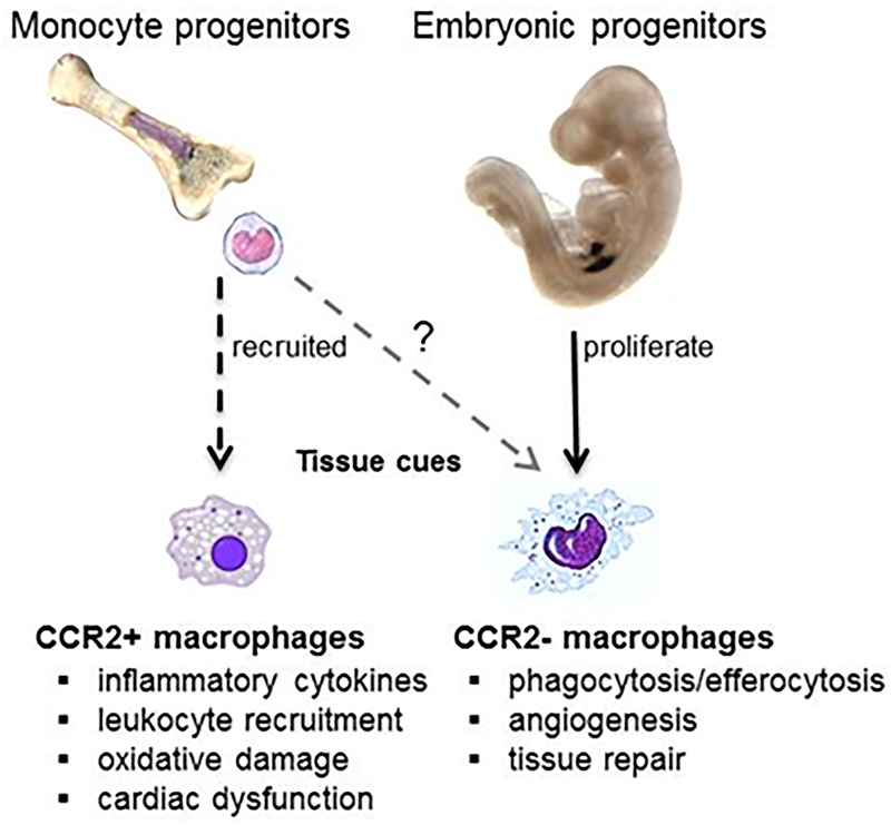
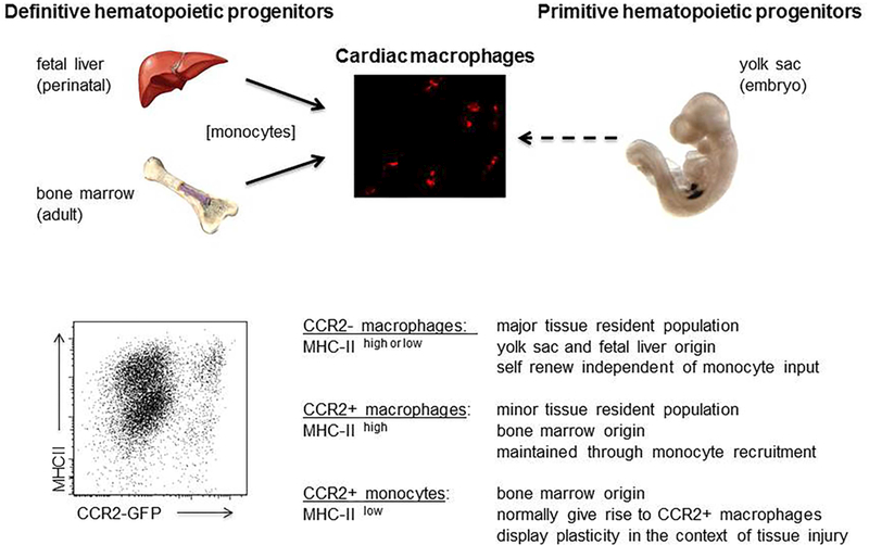
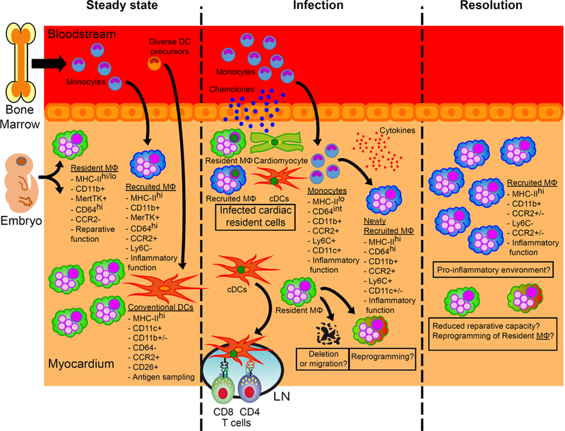
Macrophages are integral components of cardiac tissue and exert profound effects on the healthy and diseased heart. Paradigm shifting studies using advanced molecular techniques have revealed significant complexity within these macrophage populations that reside in the heart. In this final of a 4-part review series covering the macrophage in cardiovascular disease, the authors review the origins, dynamics, cell surface markers, and respective functions of each cardiac macrophage subset identified to date, including in the specific scenarios of myocarditis and after myocardial infarction. Looking ahead, a deeper understanding of the diverse and often dichotomous functions of cardiac macrophages will be essential for the development of targeted therapies to mitigate injury and orchestrate recovery of the diseased heart. Moreover, as macrophages are critical for cardiac healing, they are an emerging focus for therapeutic strategies aimed at minimizing cardiomyocyte death, ameliorating pathological cardiac remodeling, and for treating heart failure and after myocardial infarction.

Keywords: coronary angiogenesis; heart development; heart failure; macrophage; ontogeny.


Conflict of interest statement

Disclosures: The authors declare no conflicts of interest.

Figures

Central Illustration.
Central Illustration.. Myocardial injury triggers a dramatic shift in macrophage composition.
While tissue resident CCR2 macrophages expand through local proliferation, resident macrophage subsets are vastly outnumbered by recruited monocytes. Upon entering the heart, monocytes predominantly differentiate into CCR2+ macrophages with smaller contributions to other macrophage subsets (i.e., CCR2 macrophages). CCR2+ macrophages display a predominantly pro-inflammatory phenotype, while CCR2 macrophages exhibit a reparative/regenerative phenotype.
Figure 1:
Figure 1:. Macrophage heterogeneity and functions in the adult mouse heart.
Top, Schematic depicting the developmental origins of cardiac macrophages. Bottom, Flow cytometry showing macrophage populations within the adult heart during homeostasis and the mechanisms by which each population is maintained. CCR2 expression was examined by measuring GFP fluorescence in Ccr2-GFP reporter mice. Adapted from (21).
Figure 2.
Figure 2.. Macrophage dynamics and phenotype during cardiac injury.
During phase 1 or the ‘inflammatory’ phase of the injury response, Ly6chigh monocytes are the predominant monocyte/macrophage population. During phase 2, or the ‘reparative’ phase, Ly6clow macrophages are the most prevalent. Figure based on data from (7,39,40) and adapted from (40).
Figure 3.
Figure 3.. Signaling circuitry promoting monocyte and macrophage accumulation in the injured heart.
(Green pathway) Circulating DAMPs and inflammatory mediators mobilize bone marrow hematopoietic progenitors and monocytes. Monocytes enter the circulation, whereas progenitors migrate to the spleen and support splenic monocytopoiesis. Monocytes from the bone marrow and spleen eventually arrive at the injured heart. (Blue pathway) Pain and anxiety activate sympathetic pathways suppressing hematopoietic progenitor retention factors in the bone marrow leading to further progenitor cell release. (Red pathway) The injured heart directly signals to the spleen and induces splenic monocytopoiesis and hematopoietic progenitor cell proliferation by IL-1β and AngII dependent mechanisms. (Yellow pathway) Inflammatory mediators and DAMPs signal directly to recruit monocytes. Adapted from (157).
Figure 4.
Figure 4.. The role of macrophages in viral myocarditis.
In homeostasis, two ontologically different populations of macrophages (MΦ), with different functions, reside in the heart. Upon viral infection, the resident cells of the myocardium produce chemokines that promote the recruitment of monocytes, which in turn differentiate into macrophages with proinflammatory functions. However, besides the fact that their numbers recede, little is known about the role of resident macrophages during and after infection, and many questions remain (see boxes). The difficulty in distinguishing these different populations using (significantly overlapping) surface markers adds to the challenges faced in addressing these questions.

References

    1. Mann DL. Innate immunity and the failing heart: the cytokine hypothesis revisited. Circ Res 2015;116:1254–68. - PMC - PubMed
    1. Jolly SR, Kane WJ, Hook BG, Abrams GD, Kunkel SL, Lucchesi BR. Reduction of myocardial infarct size by neutrophil depletion: effect of duration of occlusion. Am Heart J 1986;112:682–90. - PubMed
    1. Horckmans M, Ring L, Duchene J et al. Neutrophils orchestrate post-myocardial infarction healing by polarizing macrophages towards a reparative phenotype. Eur Heart J 2017;38:187–197. - PubMed
    1. Litt MR, Jeremy RW, Weisman HF, Winkelstein JA, Becker LC. Neutrophil depletion limited to reperfusion reduces myocardial infarct size after 90 minutes of ischemia. Evidence for neutrophil-mediated reperfusion injury. Circulation 1989;80:1816–27. - PubMed
    1. Glaros T, Larsen M, Li L. Macrophages and fibroblasts during inflammation, tissue damage and organ injury. Front Biosci (Landmark Ed) 2009;14:3988–93. - PubMed

Publication types

MeSH terms