. 2018 Nov 30;362(6418):1064-1069.
doi: 10.1126/science.aau2818. Epub 2018 Oct 25.

Pathogen blockade of TAK1 triggers caspase-8-dependent cleavage of gasdermin D and cell death

Affiliations

Pathogen blockade of TAK1 triggers caspase-8-dependent cleavage of gasdermin D and cell death

Pontus Orning et al. Science. .

Abstract

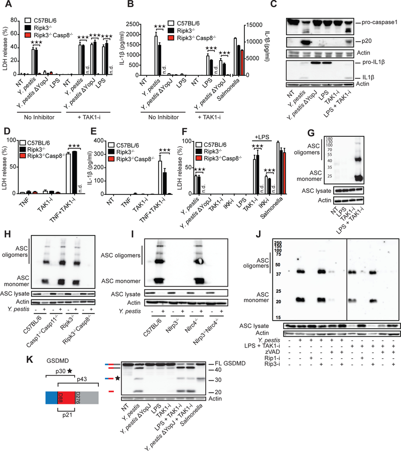
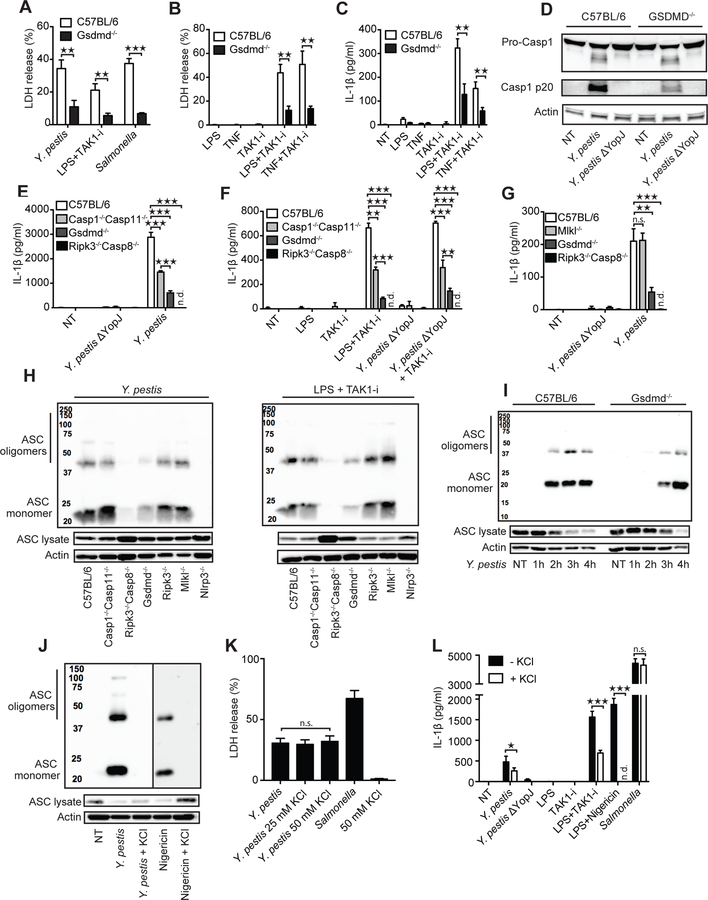
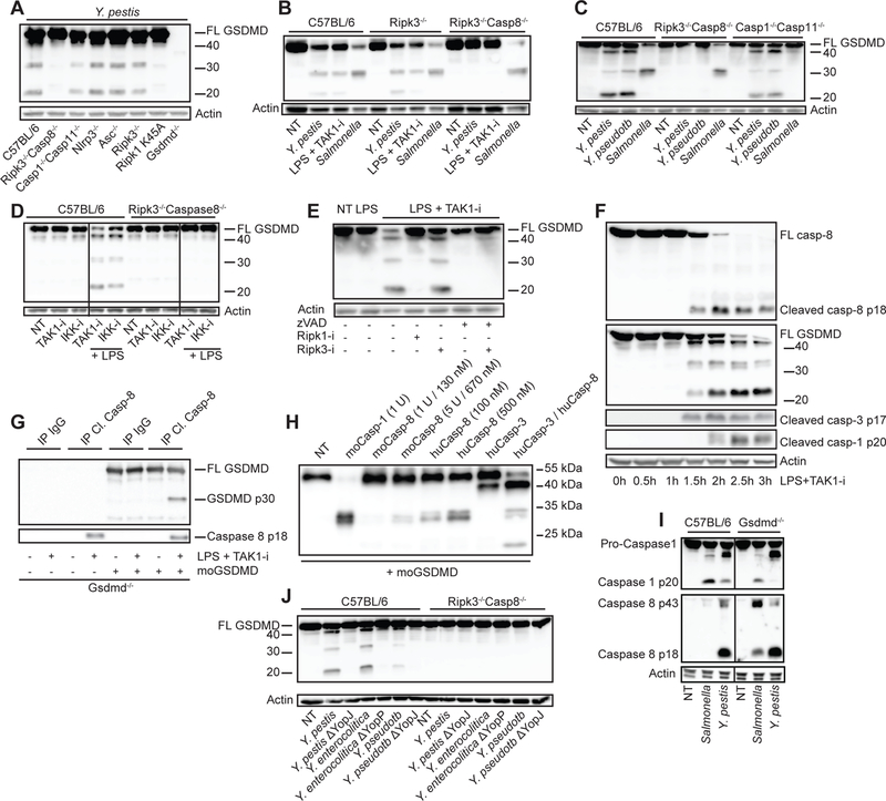
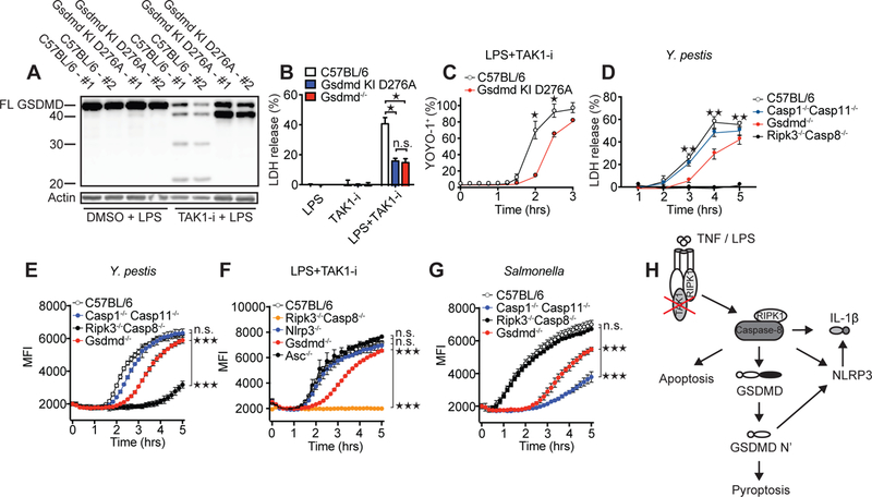
Limited proteolysis of gasdermin D (GSDMD) generates an N-terminal pore-forming fragment that controls pyroptosis in macrophages. GSDMD is processed via inflammasome-activated caspase-1 or -11. It is currently unknown whether macrophage GSDMD can be processed by other mechanisms. Here, we describe an additional pathway controlling GSDMD processing. The inhibition of TAK1 or IκB kinase (IKK) by the Yersinia effector protein YopJ elicits RIPK1- and caspase-8-dependent cleavage of GSDMD, which subsequently results in cell death. GSDMD processing also contributes to the NLRP3 inflammasome-dependent release of interleukin-1β (IL-1β). Thus, caspase-8 acts as a regulator of GSDMD-driven cell death. Furthermore, this study establishes the importance of TAK1 and IKK activity in the control of GSDMD cleavage and cytotoxicity.


Conflict of interest statement

Competing interests: BL and NK are employees of Genentech. SBB, PJG and JB are employees and shareholders of GlaxoSmithKline.

Figures

Fig. 1
Fig. 1. Inhibition of TAK1 by Yersinia YopJ or small molecules triggers cell death, inflammasome activation and GSDMD cleavage.
BMDMs from C57BL/6, Ripk3−/−, and Ripk3−/−Caspase8−/− mice were challenged with indicated stimuli in the presence or absence of TAK1 inhibitor 5z7 (TAK1-i) or IKK inhibitor (IKK-i) (A, B, D-F). Cell death was measured by LDH release after 4 h (A, D and F) and IL-1β release by ELISA after 5h (B and E). (C) C57BL/6 BMDMs were challenged with indicated stimuli for 5 h, and cell lysates plus supernatants were analyzed by immunoblot for caspase-1 and IL-1β cleavage. (G-J) Oligomerization of ASC in the inflammasome-enriched and cross-linked lysates was detected in C57BL/6 (G, J) or the indicated KO BMDMs infected or stimulated for 3 hrs with LPS + TAK1- i, with or without 1 h pretreatment with inhibitors of pan-caspase (zVAD-fmk, 25 μM), RIP1 (1 μM) or RIP3 (3 μM). (K) C57BL/6 BMDMs were treated with indicated stimuli for 3 h and cell lysates were analyzed by immunoblot for GSDMD cleavage. Illustration of GSDMD, predicted cleavage sites and fragment sizes are indicated. Data are presented as mean ± SD of triplicate wells from three or more independent experiments. n.d.: not detected. For comparisons we used t-test. *p<0.05, **p<0.01, ***p<0.001.
Fig. 2
Fig. 2. GSDMD regulates cell death and inflammasome activation in response to TAK1 inhibition.
(A-G) The indicated BMDMs were challenged with Y. pestis or a mutant lacking YopJ, or S. Typhimurium, or stimulated with indicated ligands, TAK1-i, or ligands + TAK1-i. Cell death was measured by LDH release after 3 h (A and B), and IL-1β release after 5 h (C, E-G), or caspase-1 cleavage after 5h (D). (H-J) Oligomerization of ASC in the inflammasome-enriched and cross-linked lysates was detected in BMDMs after Y. pestis or LPS + TAK1-I challenge after the indicated time points (I) or 3 h. (J-L) BMDMs treated with Y. pestis, S. Typhimurium, or nigericin + LPS in the presence or absence of 50 mM (J-L) or 25 mM (K) KCl, and oligomerization of ASC was detected by immunoblot, cell death was measured by LDH release, or IL-1β by ELISA. Data are presented as mean ± SD of triplicate wells from three or more independent experiments. n.d.: not detected. For comparisons between two groups we used t-test, for more than two groups we used ANOVA. *p<0.05, **p<0.01, ***p<0.001.
Fig. 3
Fig. 3. Caspase-8 dependent GSDMD processing with little contribution by caspase-1 and -11.
BMDMs from the indicated mouse strains were challenged with the bacteria Y. pestis, Y. pseudotuberculosis, Y. enterocolitica, their YopJ (YopP in Y.e.) mutants, or S. Typhimurium, or treated with LPS + TAK1-i. Cell lysates were harvested after 3–4 h unless otherwise stated and analyzed by immunoblots for GSDMD cleavage (A-F, and J) or caspase-1, caspase-3 and caspase-8 cleavage (F, I). (D) BMDMs from C57BL/6 or Ripk3−/−Casp8−/− mice were treated with IKK-i and TAK1-i in the absence or presence of LPS. (E) BMDMs were pretreated with pan-caspase inhibitor zVAD-fmk, RIPK1-i, or RIPK3-i for 1 h before being treated with LPS+TAK1-i. (G) Gsdmd−/− BMDMs were treated with LPS+TAK1-i for 3 h and then immunoprecipitated with cleaved caspase-8 antibody. Immunoprecipitates (G) or recombinant caspases (H) were incubated with recombinant mouse GSDMD at 37°C for 1 h in a protein cleavage buffer, before being analyzed by immunoblots. Figures are representative of three or more independent experiments.
Fig. 4
Fig. 4. The GSDMD Asp276 residue is critical for GSDMD processing and for regulation of cell death downstream of TAK1 inhibition.
(A) Immunoblot of GSDMD cleavage in C57BL/6 or Gsdmd D276A mutant BMDMs after treatment with LPS and LPS + TAK1-i. Cell death as measured by LDH release (B and D) or by % YOYO-1+ cells (C) after challenge of indicated genotypes with LPS + TAK1-i or Y. pestis. (E-G) Cell death as measured by entry of EthD-1 into the cell. Samples were read every 10 min at 530 nm excitation, 645 nm emission wavelength. BMDMs from the indicated genotypes were treated with Y. pestis (E), LPS + TAK1-i (F), or S. Typhimurium (G). Infections in E, G are without gentamicin. For (E-G) statistics were performed on the area under the curve (AUC) for the whole time course. (H) Proposed model of TAK1-inhibition-induced caspase-8 activation and cell death. Data are presented as mean ± SD of triplicate wells from two (A-C) or three or more (D-G) independent experiments. n.d. not detected. For comparisons between two groups we used t-test, for more than two groups we used ANOVA. *p<0.05, **p<0.01, ***p<0.001.

References

    1. Sborgi L et al., GSDMD membrane pore formation constitutes the mechanism of pyroptotic cell death. EMBO J 35, 1766–1778 (2016). - PMC - PubMed
    1. Liu X et al., Inflammasome-activated gasdermin D causes pyroptosis by forming membrane pores. Nature 535, 153–158 (2016). - PMC - PubMed
    1. Kovacs SB, Miao EA, Gasdermins: Effectors of Pyroptosis. Trends Cell Biol 27, 673–684 (2017). - PMC - PubMed
    1. Shi J, Gao W, Shao F, Pyroptosis: Gasdermin-Mediated Programmed Necrotic Cell Death. Trends Biochem. Sci 42 (2017), pp. 245–254. - PubMed
    1. Shi J et al., Cleavage of GSDMD by inflammatory caspases determines pyroptotic cell death. Nature 526, 660–665 (2015). - PubMed

Publication types

MeSH terms