. 2018 Nov 26;7(11):bio038661.
doi: 10.1242/bio.038661.

Plag1 and Plagl2 have overlapping and distinct functions in telencephalic development

Affiliations

Plag1 and Plagl2 have overlapping and distinct functions in telencephalic development

Lata Adnani et al. Biol Open. .

Abstract

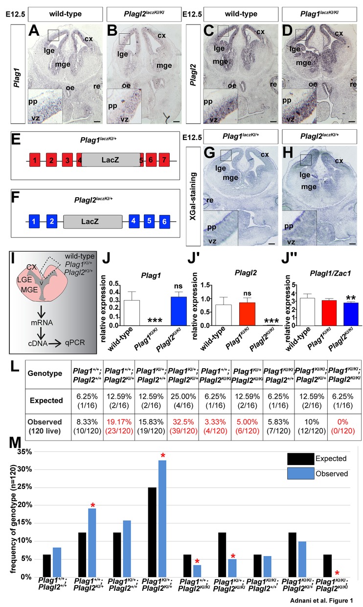
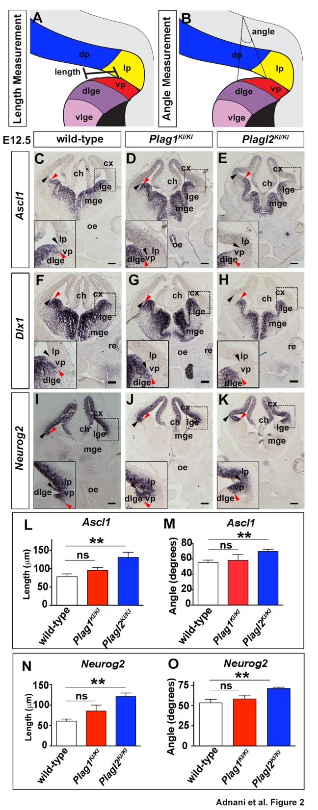
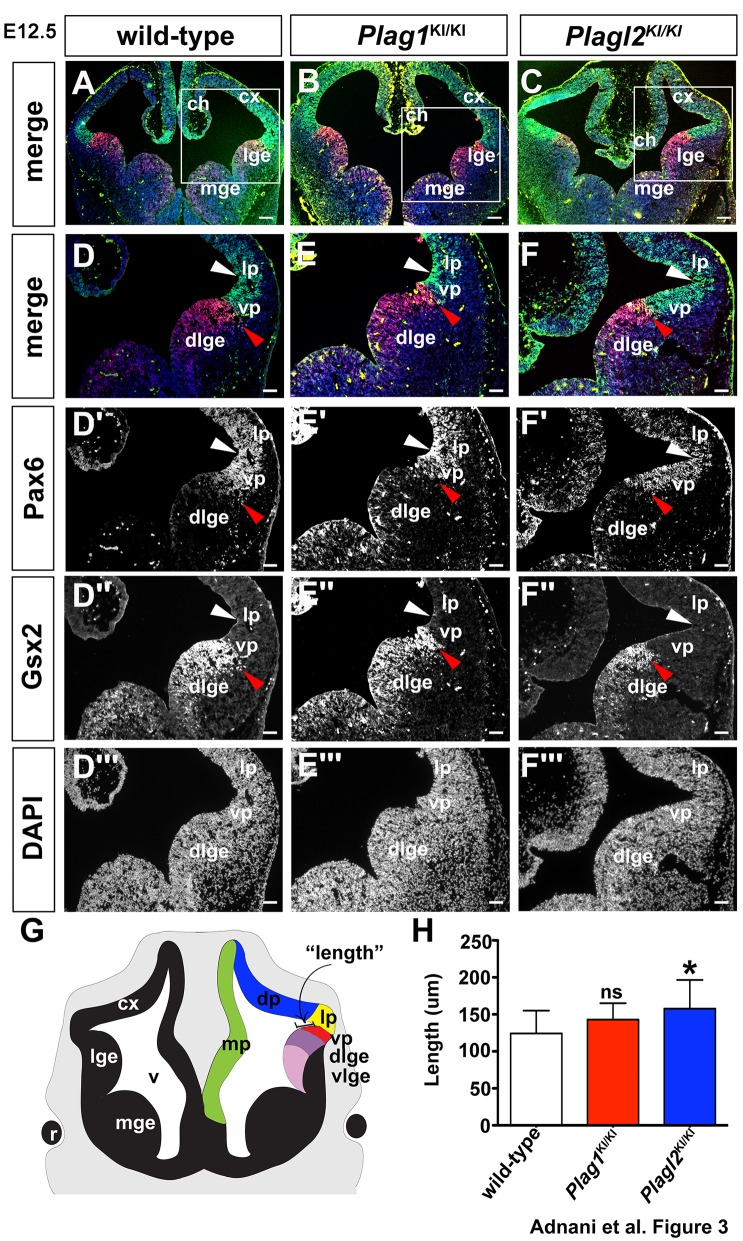
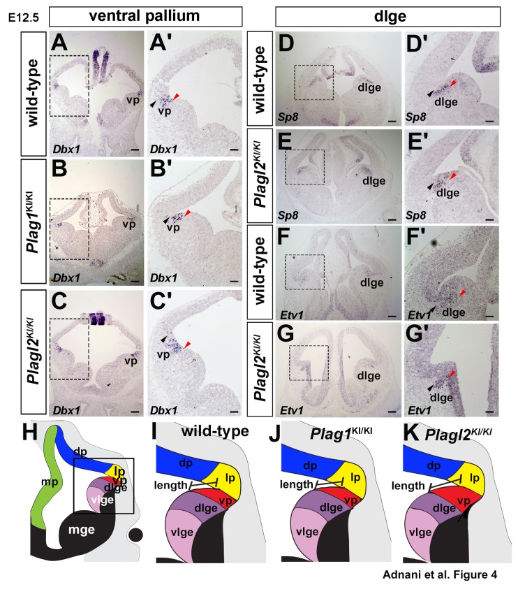
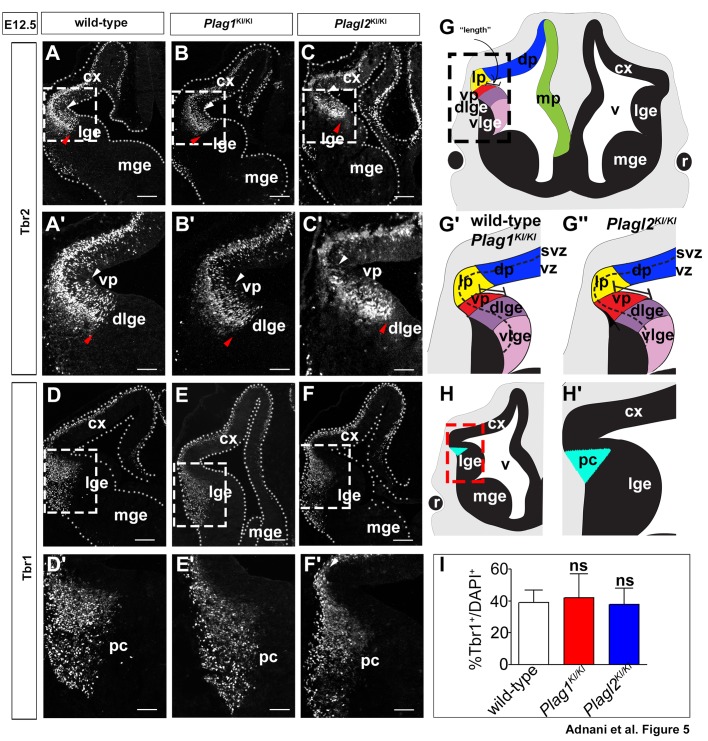
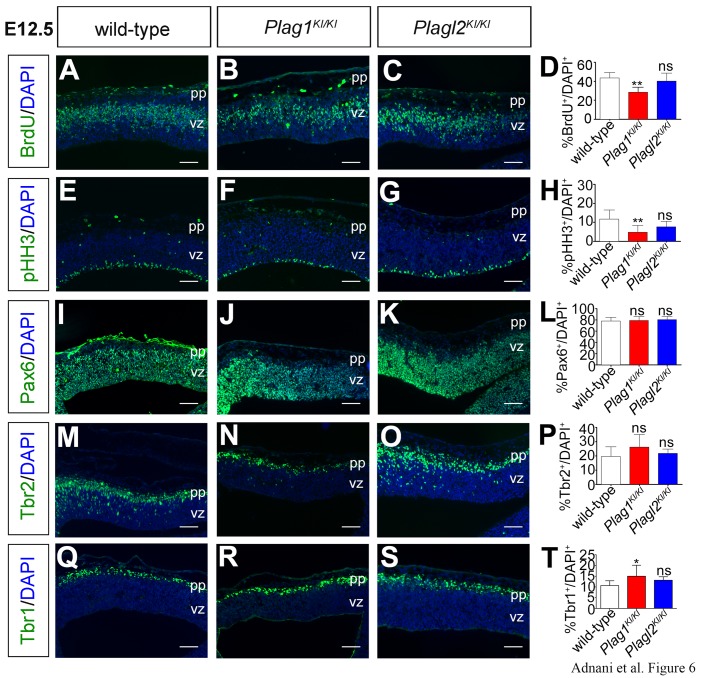
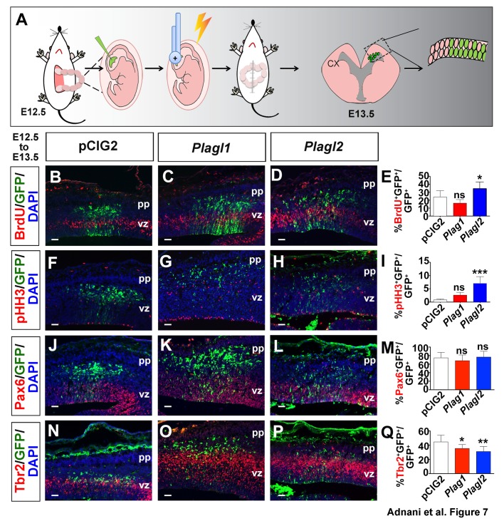
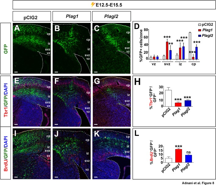
The Plag gene family has three members; Plagl1/Zac1, which is a tumor suppressor gene, and Plag1 and Plagl2, which are proto-oncogenes. All three genes are known to be expressed in embryonic neural progenitors, and Zac1 regulates proliferation, neuronal differentiation and migration in the developing neocortex. Here we examined the functions of Plag1 and Plagl2 in neocortical development. We first attempted, and were unable to generate, E12.5 Plag1;Plagl2 double mutants, indicating that at least one Plag1 or Plagl2 gene copy is required for embryonic survival. We therefore focused on single mutants, revealing a telencephalic patterning defect in E12.5 Plagl2 mutants and a proliferation/differentiation defect in Plag1 mutant neocortices. Specifically, the ventral pallium, a dorsal telencephalic territory, expands into the ventral telencephalon in Plagl2 mutants. In contrast, Plag1 mutants develop normal regional territories, but neocortical progenitors proliferate less and instead produce more neurons. Finally, in gain-of-function studies, both Plag1 and Plagl2 reduce neurogenesis and increase BrdU-uptake, indicative of enhanced proliferation, but while Plagl2 effects on proliferation are more immediate, Plag1 effects are delayed. Taken together, we found that the Plag proto-oncogenes genes are essential regulators of neocortical development and although Plag1 and Plagl2 functions are similar, they do not entirely overlap. This article has an associated First Person interview with the first author of the paper.

Keywords: Neocortical development; Neural progenitor proliferation; Neurogenesis; Plag gene family; Telencephalic patterning; Zinc finger transcription factors.


Conflict of interest statement

Competing interestsThe authors declare no competing or financial interests.

Figures

Fig. 1.
Fig. 1.
Plag1 and Plagl2 have similar patterns of telencephalic gene expression and function redundantly to regulate embryonic development. (A–D) Expression of Plag1 (A,B) and Plagl2 (C,D) in E12.5 wild-type (A,C), Plagl2lacZKI/KI (B) and Plag1lacZKI/KI (D) whole heads. (E,F) Schematic representations of genetic mutations in Plag1lacZKI/KI (E) and Plagl2lacZKI/KI (F) mice (G,H) X-gal staining of E12.5 PlagllacZKI/+ (G) and Plagl2lacZKI/+ (H) brains. (I–J″) Schematic representation of RT-qPCR experiment (I). Analysis of Plag1 (J), Plagl2 (J′) and Plagl1/Zac1 (J″) transcript levels in E12.5 wild-type, Plag1KI/KI and Plagl2KI/KI cortices. ns, not significant; *P<0.05, **P<0.01, and ***P<0.005. (L,M) Punette square analysis of the ratios of genotypes acquired from Plag1KI/+;Plagl2KI/+ heterozygous intercrosses (L). Graphical representation of the expected (black bars) and observed (blue bars) numbers of embryos with each genotype (M). cx, neocortex; lge, lateral ganglionic eminence; mge, medial ganglionic eminence; oe, olfactory epithelium; pp, preplate; re, retina; vz, ventricular zone. Scale bars: 250 μm.
Fig. 2.
Fig. 2.
Plag1 and Plagl2 are required to pattern the embryonic telencephalon. (A,B) Schematic representations of length (A) and angle (B) measurements of the ventral pallium, extending from the corticostriatal angle to the gene expression border. (C–K) Expression of Ascl1 (C–E), Dlx1 (F–H) and Neurog2 (I–K) in E12.5 wild-type (C,F,I), Plag1KI/KI (D,G,J) and Plagl2KI/KI (E,H,K) brains. Black arrowheads mark the corticostriatal angle and red arrowheads mark the ventral pallial gene expression limit. (L–O) Quantification of the length (L,N) and angle (M,O) of the ventral pallium based on the expression of Ascl1 (L,M), and Neurog2 (N,O). Error bars are s.e.m. ns, not significant; *P<0.05, **P<0.01, and ***P<0.005. ch, cortical hem; cx, neocortex; dlge, dorsal lateral ganglionic eminence; dp, dorsal pallium; lge, lateral ganglionic eminence; lp, lateral pallium; mge, medial ganglionic eminence; mp, medial pallium; oe, olfactory epithelium; re, retina; vlge, ventral lateral ganglionic eminence; vp, ventral pallium. Scale bars: 250 μm.
Fig. 3.
Fig. 3.
Plagl2 is required to maintain the pallial-subpallial boundary in the developing telencephalon. (A–F) Expression of Pax6 (green), Gsh2 (red) and DAPI (blue) in E12.5 wild-type (A,D–D‴), Plag1KI/KI (B,E–E‴) and Plagl2KI/KI (C,F–F‴) brains. (D′–F‴) Higher magnification images of Pax6 (D′–F′), Gsh2 (D″–F″) and DAPI (D‴–F‴) in E12.5 wild-type (D′–D‴), Plag1KI/KI (E′–E‴) and Plagl2KI/KI (F′–F‴) brains. White arrowheads mark the corticostriatal angle. Red arrowheads mark the ventral pallium-dlge boundary. (G) Schematic illustration of the length measurement of the ventral pallium. (H) Quantification of the length of the ventral pallium. Error bars are s.e.m. ns, not significant; *P<0.05, **P<0.01, and ***P<0.005. ch, cortical hem; cx, neocortex; dlge, dorsal lateral ganglionic eminence; dp, dorsal pallium; lge, lateral ganglionic eminence; lp, lateral pallium; mge, medial ganglionic eminence; mp, medial pallium; oe, olfactory epithelium; re, retina; vlge, ventral lateral ganglionic eminence; vp, ventral pallium. Scale bars: 250 μm.
Fig. 4.
Fig. 4.
Plagl2 is required to regulate the ventral pallium boundary in the developing telencephalon. (A–C′) Expression of the ventral pallial gene, Dbx1, in E12.5 wild-type (A,A′), Plag1KI/KI (B,B′) and Plagl2KI/KI (C,C′) telencephalons. (D–G′) Expressions of the dlge markers, Sp8 (D–E′) and Etv1 (F–G′) in E12.5 wild-type (D,D′,F,F′) and Plagl2KI/KI (E,E′,G,G′) telencephalons. Black arrowheads mark the corticostriatal angle. Red arrowheads mark the dorsal limit of gene expression. (H–K) Schematic illustration of the pallial-subpallial boundary between the ventral pallium and dlge in E12.5 wild-type (H,I), Plag1KI/KI (J) and Plagl2KI/KI (K), showing positioning defects in the Plagl2KI/KI brains. dlge, dorsal lateral ganglionic eminence; vp, ventral pallium. Scale bars: 250 μm.
Fig. 5.
Fig. 5.
Tbr2 expression in intermediate neuronal progenitors in the ventral pallium shifts ventrally in Plagl2 mutants. (A–C′) Tbr2 expression in E12.5 wild-type (A,A′), Plag1KI/KI (B,B′) and Plagl2KI/KI (C,C′) telencephalons. (D–F′) Tbr1 expression in E12.5 wild-type (D,D′), Plag1KI/KI (E,E′) and Plagl2KI/KI (F,F′) telencephalons. White arrowheads mark the corticostriatal angle. Red arrowheads mark the ventral limit of high dorsal gene expression. (G–G″) Schematic illustration of ventral pallial domain in wild-type (G,Gʹ) and Plagl2KI/KI (G″) telencephalons. (H–Hʹ) Schematic illustration of the Tbr1 piriform cortex (blue) that was used for quantification. (I) Quantification of the Tbr1+/DAPI+ cells in the presumptive piriform cortex. Error bars are s.e.m. ns, not significant. cx, neocortex; dlge, dorsal lateral ganglionic eminence; lge, lateral ganglionic eminence; mge, medial ganglionic eminence; pc, piriform cortex; vp, ventral pallium. Scale bars: 250 μm.
Fig. 6.
Fig. 6.
Plag1 is required to regulate proliferation in the early embryonic telencephalon. (A–T) Analysis of the expression of BrdU (A–C), pHH3 (E–G), Pax6 (I–K), Tbr2 (M–O) and Tbr1 (Q–S) in E12.5 wild-type (A,E,I,M,Q), Plag1KI/KI (B,F,J,N,R) and Plagl2KI/KI (C,G,K,O,S) cortices. Quantification of the percentage of DAPI+ cells expressing BrdU (D), pHH3 (H), Pax6 (L), Tbr2 (P) and Tbr1 (T). Error bars are s.e.m. ns, not significant; *P<0.05, **P<0.01, and ***P<0.005. pp, preplate; vz, ventricular zone. Scale bars: 125 μm.
Fig. 7.
Fig. 7.
Plagl2 is sufficient to alter the proliferation of neocortical progenitors within 24 h post-electroporation. (A) Schematic representation of gain-of-function experiment using in utero electroporation. (B–Q) E12.5 to E13.5 electroporations of pCIG2 (B,F,J,N), Plag1 (C,G,K,O) and Plagl2 (D,H,L,P) analyzed for the expression of BrdU (B–D), pHH3 (F–H), Pax6 (J–L), Tbr2 (N–P). Quantitation of the ratios of GFP+ cells that are BrdU+ (E), pHH3+ (I), Pax6+ (M) and Tbr2+ (Q). Error bars are s.e.m. ns, not significant; *P<0.05, **P<0.01, and ***P<0.005. pp, preplate; vz, ventricular zone. Scale bars: 125 μm.
Fig. 8.
Fig. 8.
Plag1 and Plagl2 are sufficient to induce migration defects, and alter proliferation and differentiation when misexpressed in the neocortex 72 h post electroporation. (A–L) E12.5 to E15.5 electroporations of pCIG2 (A,E,I), Plag1 (B,F,J) and Plagl2 (C,G,K) analyzed for the expression of GFP in different zones (A–D), and the co-expression of GFP with Tbr1 (E–H) and BrdU (I–L). Quantitation of the ratios of GFP+ cells that are in each zone (D), Tbr1+ (H) and BrdU+ (L). Error bars are s.e.m. ns, not significant; *P<0.05, **P<0.01, and ***P<0.005. cp, cortical plate; iz, intermediate zone; svz, subventricular zone; vz, ventricular zone. Scale bars: 125 μm.

References

    1. Abdollahi A. (2007). LOT1 (ZAC1/PLAGL1) and its family members: mechanisms and functions. J. Cell. Physiol. 210, 16-25. 10.1002/jcp.20835 - DOI - PubMed
    1. Abdollahi A., Godwin A. K., Miller P. D., Getts L. A., Schultz D. C., Taguchi T., Testa J. R. and Hamilton T. C. (1997a). Identification of a gene containing zinc-finger motifs based on lost expression in malignantly transformed rat ovarian surface epithelial cells. Cancer Res. 57, 2029-2034. - PubMed
    1. Abdollahi A., Roberts D., Godwin A. K., Schultz D. C., Sonoda G., Testa J. R. and Hamilton T. C. (1997b). Identification of a zinc-finger gene at 6q25: a chromosomal region implicated in development of many solid tumors. Oncogene 14, 1973-1979. 10.1038/sj.onc.1201034 - DOI - PubMed
    1. Adnani L., Langevin L. M., Gautier E., Dixit R., Parsons K., Li S., Kaushik G., Wilkinson G., Wilson R., Childs S. et al. (2015). Zac1 regulates the differentiation and migration of neocortical neurons via Pac1. J. Neurosci. 35, 13430-13447. 10.1523/JNEUROSCI.0777-15.2015 - DOI - PMC - PubMed
    1. Adnani L., Han S., Li S., Mattar P. and Schuurmans C. (2018). Mechanisms of cortical differentiation. Int. Rev. Cell Mol. Biol. 336, 223-320. 10.1016/bs.ircmb.2017.07.005 - DOI - PubMed