. 2019 Mar;104(3):587-598.
doi: 10.3324/haematol.2018.203166. Epub 2018 Oct 25.

Unraveling the effect of silent, intronic and missense mutations on VWF splicing: contribution of next generation sequencing in the study of mRNA

Affiliations

Unraveling the effect of silent, intronic and missense mutations on VWF splicing: contribution of next generation sequencing in the study of mRNA

Nina Borràs et al. Haematologica. 2019 Mar.

Abstract

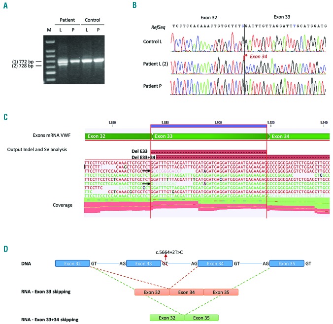
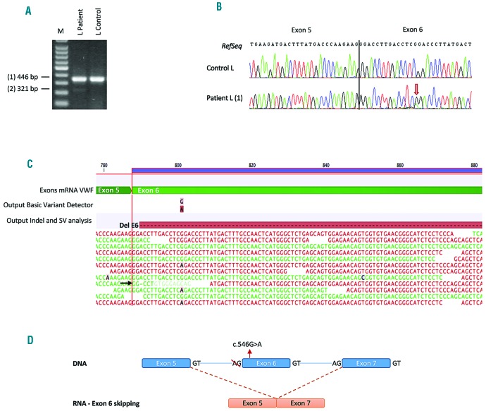
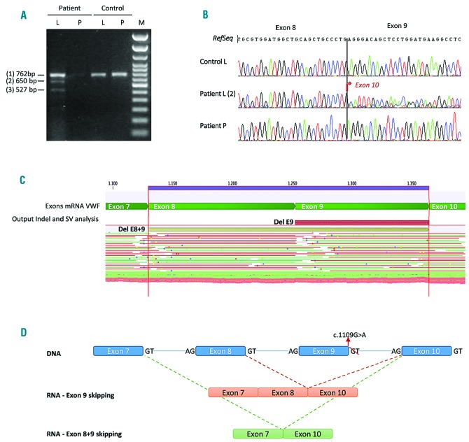
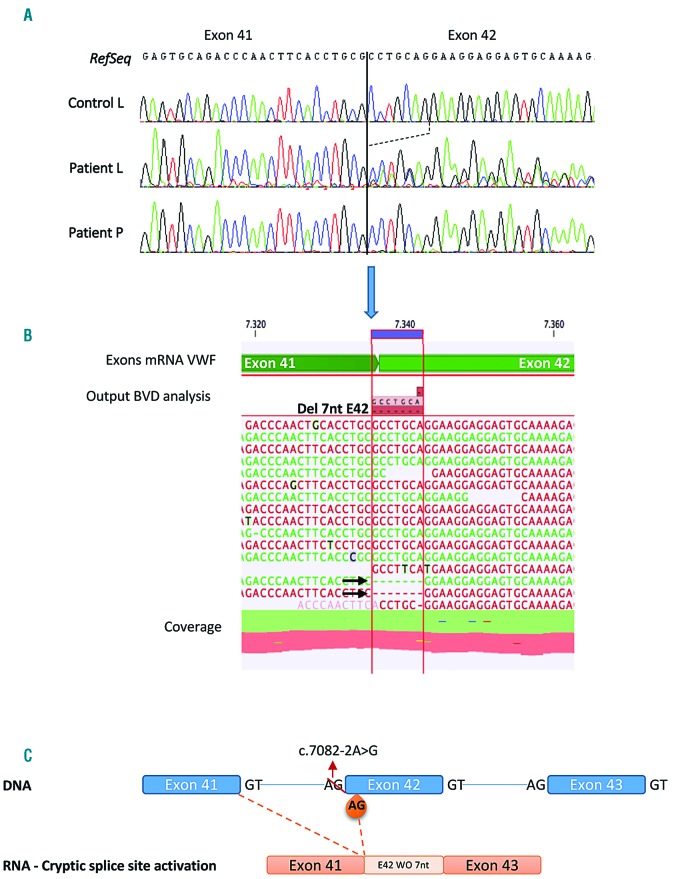
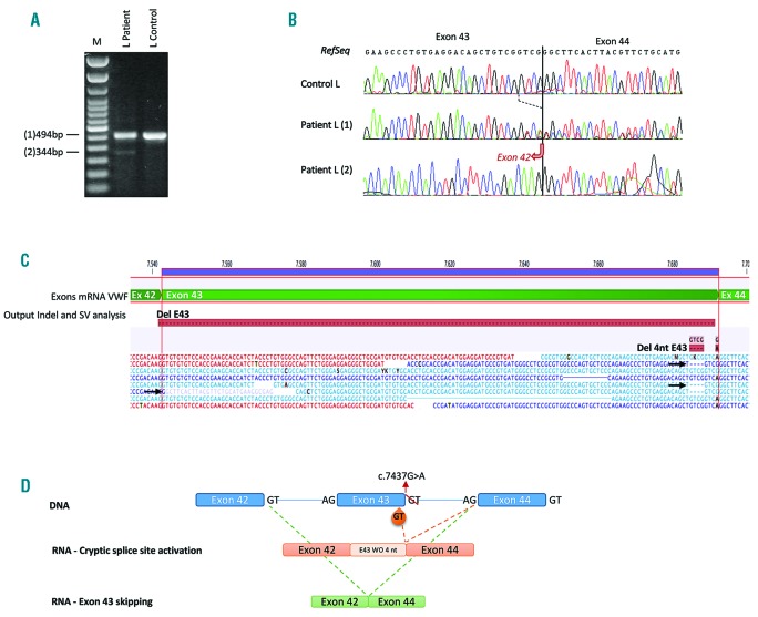
Large studies in von Willebrand disease patients, including Spanish and Portuguese registries, led to the identification of >250 different mutations. It is a challenge to determine the pathogenic effect of potential splice site mutations on VWF mRNA. This study aimed to elucidate the true effects of 18 mutations on VWF mRNA processing, investigate the contribution of next-generation sequencing to in vivo mRNA study in von Willebrand disease, and compare the findings with in silico prediction. RNA extracted from patient platelets and leukocytes was amplified by RT-PCR and sequenced using Sanger and next generation sequencing techniques. Eight mutations affected VWF splicing: c.1533+1G>A, c.5664+2T>C and c.546G>A (p.=) prompted exon skipping; c.3223-7_3236dup and c.7082-2A>G resulted in activation of cryptic sites; c.3379+1G>A and c.7437G>A) demonstrated both molecular pathogenic mechanisms simultaneously; and the p.Cys370Tyr missense mutation generated two aberrant transcripts. Of note, the complete effect of three mutations was provided by next generation sequencing alone because of low expression of the aberrant transcripts. In the remaining 10 mutations, no effect was elucidated in the experiments. However, the differential findings obtained in platelets and leukocytes provided substantial evidence that four of these would have an effect on VWF levels. In this first report using next generation sequencing technology to unravel the effects of VWF mutations on splicing, the technique yielded valuable information. Our data bring to light the importance of studying the effect of synonymous and missense mutations on VWF splicing to improve the current knowledge of the molecular mechanisms behind von Willebrand disease. clinicaltrials.gov identifier:02869074.


Figures

Figure 1.
Figure 1.
Analysis of the c.5664+2T>C mutation in patient UMP03. A) RT-PCR products amplified with primers located in exons 30 and 35 in leukocyte (L) and platelet (P) RNA, separated on 1% agarose gel. B) Traditional Sanger sequencing of PCR products from patient leukocytes (L2) and platelets (P), and control leukocytes (L). Analysis of the band 2 from patient leukocyte on agarose gel demonstrates exon 33 skipping. In platelets, only the allele without c.5664+2T>C mutation could be amplified. C) NGS of PCR products from leukocytes showed exon 33 skipping, indicated by arrows depict aberrant transcripts. However, exon 33-34 skipping was also detected, but in a really low of transcripts. D) Schematic representation of the mutation in genomic DNA and its effect on the VWF mRNA sequence. M indicates a 100-bp DNA ladder.
Figure 2.
Figure 2.
Analysis of the c.546G>A mutation, located at nucleotide 14 from the beginning of exon 6, in patient UMP08. A) RT-PCR products amplified with primers located in exons 4 and 7 in leukocyte RNA (L), separated on 1% agarose gel. B) Traditional Sanger sequencing of PCR product from patient L (1) showed the single nucleotide change. C) NGS of PCR products from leukocytes identified the single nucleotide variant, as well as exon 6 skipping. Arrows show aberrant transcripts. D) Schematic representation of the mutation in genomic DNA and its effect on the VWF mRNA sequence. M indicates a 100-bp DNA ladder.
Figure 3.
Figure 3.
Analysis of the c.1109G>A (p.Cys370Tyr) mutation in patient UMP10, located in the last nucleotide of exon 9. A) RT-PCR products amplified with primers located in exons 6 and 12 in leukocyte (L) and platelet (P) RNA, separated on 1% agarose gel. B) Traditional Sanger sequencing of PCR product from patient L (2) showed exon 9 skipping. The L (3) band could not be purified in the agarose gel due to low concentration, thus it was not analyzed by Sanger. In platelets, only the non-mutated allele could be amplified. C) NGS of PCR products from leukocytes identified transcripts lacking exon 9, as well as transcripts lacking exons 8 and 9. D) Schematic representation of the mutation in genomic DNA and its effect on the VWF mRNA sequence. M indicates a 100-bp DNA ladder.
Figure 4.
Figure 4.
Analysis of the c.7082-2A>G mutation in patient UMP12. Agarose gel electrophoresis results of RT-PCR amplification of exon 38 to 43 using RNA from leukocytes and platelets were the same as those of healthy controls (data not shown). A) Traditional Sanger sequencing of PCR product from patient leukocyte (L) demonstrate activation of a cryptic splice site 7 nucleotides downstream of the native splice site within exon 42. In patient platelet (P), only the allele carrying the p.Leu150Pro mutation could be amplified. B) NGS of PCR products from leukocytes showed deletion of the 7 initial nucleotides of exon 42. Arrows show aberrant transcripts. C) Schematic representation of the mutation in genomic DNA and its effect on the VWF mRNA sequence. WO: without.
Figure 5.
Figure 5.
Analysis of the c.7437G>A (p.=) mutation in patient UMP14, located in the last nucleotide of exon 43. A) RT-PCR products amplified with primers located in exons 41 and 45 in leukocyte RNA (L), separated on 1% agarose gel. B) Traditional Sanger sequencing of PCR product from patient leukocyte (L) shows two aberrant transcripts: activation of a cryptic splice site - 4 nucleotides upstream to WT-DSS - in exon 43 (1), and exon 43 skipping (2). C) NGS of PCR products gave the same results than Sanger sequencing. D) Schematic representation of the mutation in genomic DNA and its effect on the VWF mRNA sequence. M indicates a 100-bp DNA ladder. WO: without.

Similar articles

Cited by

References

    1. Rodeghiero F, Castaman G, Dini E. Epidemiological investigation of the prevalence of von Willebrand’s disease. Blood. 1987;69(2):454–459. - PubMed
    1. Batlle J, Perez-Rodriguez A, Corrales I, et al. Molecular and clinical profile of von Willebrand disease in Spain (PCM-EVW-ES): Proposal for a new diagnostic paradigm. Thromb Haemost. 2016;115(1):40–50. - PubMed
    1. Borras N, Batlle J, Perez-Rodriguez A, et al. Molecular and clinical profile of von Willebrand disease in Spain (PCM-EVW-ES): comprehensive genetic analysis by next-generation sequencing of 480 patients. Haematologica. 2017;102(12): 2005–2014. - PMC - PubMed
    1. Fidalgo T, Salvado R, Corrales I, et al. Genotype-phenotype correlation in a cohort of Portuguese patients comprising the entire spectrum of VWD types: impact of NGS. Thromb Haemost. 2016;116(1):17–31. - PubMed
    1. Richards S, Aziz N, Bale S, et al. Standards and guidelines for the interpretation of sequence variants: a joint consensus recommendation of the American College of Medical Genetics and Genomics and the Association for Molecular Pathology. Genet Med. 2015;17(5):405–424. - PMC - PubMed

Publication types