. 2019 Jan 7;218(1):234-250.
doi: 10.1083/jcb.201806094. Epub 2018 Oct 25.

Rsp5 Ubiquitin ligase-mediated quality control system clears membrane proteins mistargeted to the vacuole membrane

Affiliations

Rsp5 Ubiquitin ligase-mediated quality control system clears membrane proteins mistargeted to the vacuole membrane

Richa Sardana et al. J Cell Biol. .

Abstract

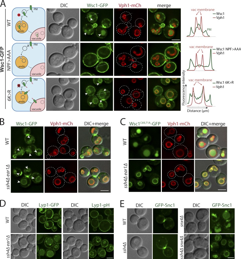
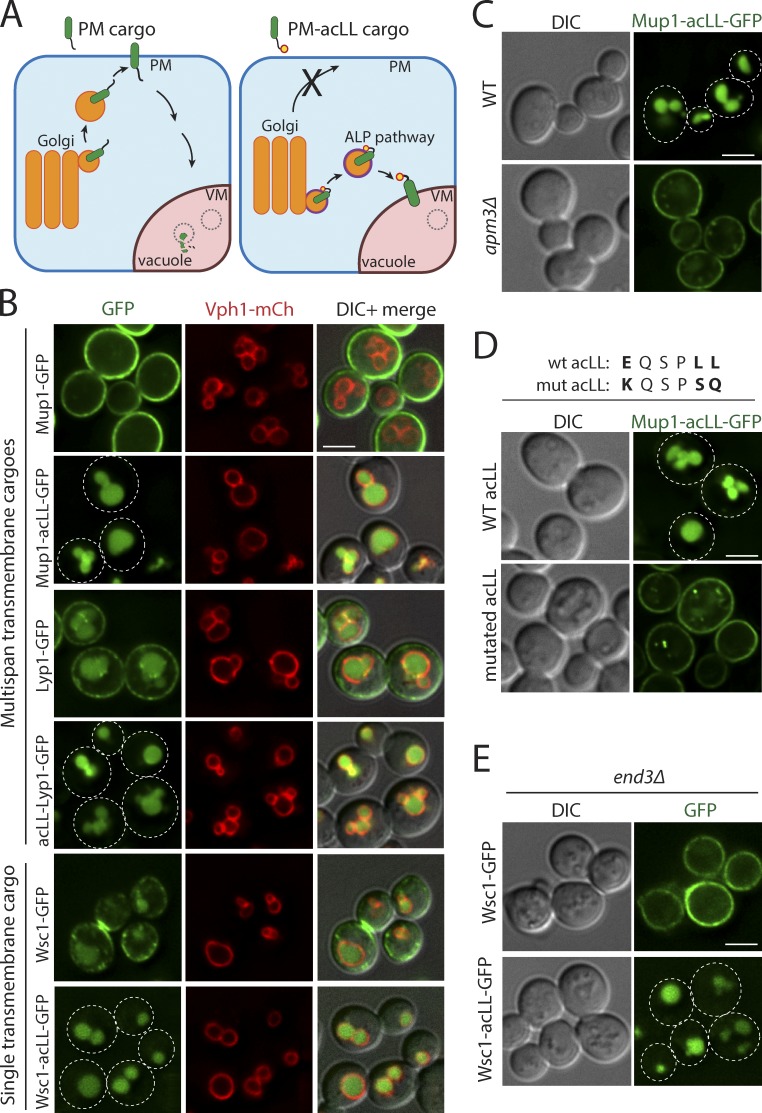
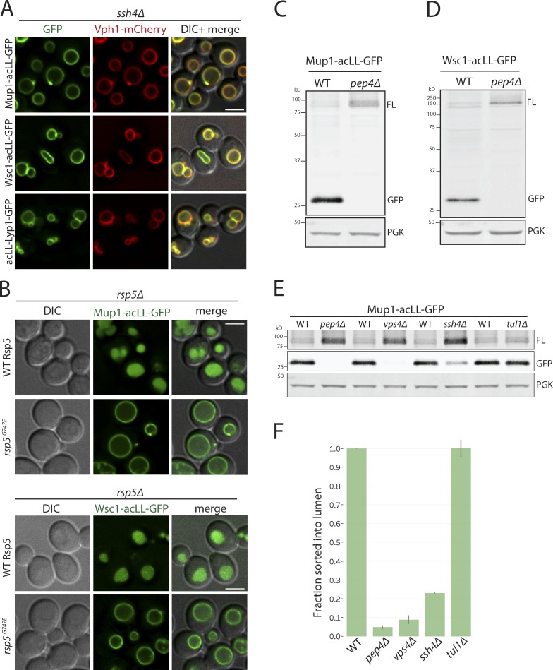
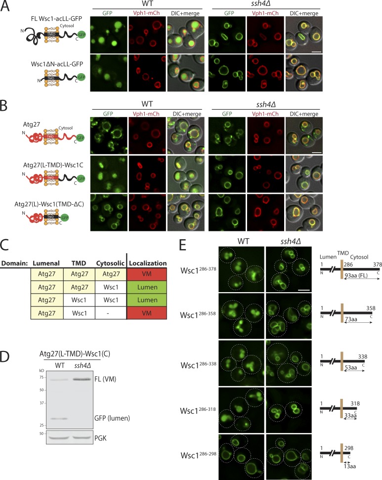
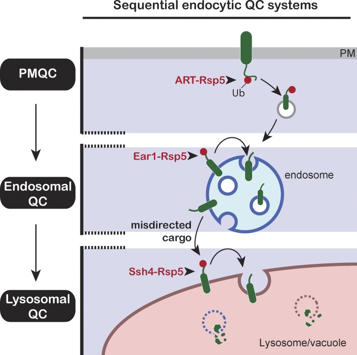
Maintenance of organelle identity is profoundly dependent on the coordination between correct targeting of proteins and removal of mistargeted and damaged proteins. This task is mediated by organelle-specific protein quality control (QC) systems. In yeast, the endocytosis and QC of most plasma membrane (PM) proteins requires the Rsp5 ubiquitin ligase and ART adaptor network. We show that intracellular adaptors of Rsp5, Ear1, and Ssh4 mediate recognition and vacuolar degradation of PM proteins that escape or bypass PM QC systems. This second tier of surveillance helps to maintain cell integrity upon heat stress and protects from proteotoxicity. To understand the mechanism of the recognition of aberrant PM cargos by Ssh4-Rsp5, we mistarget multiple PM proteins de novo to the vacuolar membrane. We found that Ssh4-Rsp5 can target and ubiquitinate multiple lysines within a restricted distance from the membrane, providing a fail-safe mechanism for a diverse cargo repertoire. The mistargeting or misfolding of PM proteins likely exposes these lysines or shifts them into the "ubiquitination zone" accessible to the Ssh4-Rsp5 complex.


Figures

Figure 1.
Figure 1.
Intracellular Rsp5 adaptors are required for vacuolar sorting of multiple endocytosed PM cargos. (A) Live-cell fluorescence and differential interference contrast (DIC) imaging of a GFP-tagged cell wall integrity sensor, Wsc1, and mutants lacking the Sla1 recognition motif (NPF>AAA) or all cytosolic lysines (6K>R) in WT yeast cells also expressing an mCherry-tagged VM marker, Vph1, a subunit of the vacuolar ATPase V0 domain. The arrowheads point to the vacuolar lumen signal for WT Wsc1 and the VM signal for Wsc16K>R mutant, respectively. The dashed line indicated on a representative cell for each condition was used to build a line-scan fluorescence profile using ImageJ. Schematic describing the localization phenotype for each condition is illustrated on the left. (B) WT and ssh4Δ ear1Δ mutant cells expressing Wsc1-GFP. The arrowheads point to the vacuolar lumen signal in WT cells and the VM signal in ssh4Δ ear1Δ mutant, respectively. (C) WT or ssh4Δ ear1Δ mutant yeast cells expressing clustering defective, constitutively endocytosed Wsc1C69, 71A-GFP. (D) WT or ssh4Δ ear1Δ double-mutant yeast cells expressing Lyp1 fused to GFP (Lyp1-GFP) or a pH-sensitive pHluorin (Lyp1-pHluorin). (E) Fluorescence microscopy of GFP-tagged v-SNARE Snc1 in WT, ssh4Δ, snx4Δ single mutants and ssh4Δ snx4Δ double mutant. Scale bars: 2.5 µm.
Figure 2.
Figure 2.
Addition of acLL motif constitutively mistargets PM proteins to the vacuole. (A) The strategy for mistargeting PM cargos to the vacuole. PM cargos are normally packaged into secretory vesicles at the Golgi that fuse with the PM (left). Addition of a 6-aa acidic dileucine (EQSPLL) or acLL motif (indicated by a yellow dot with a red border) to the cytosolic tails of PM proteins (PM-acLL cargo), engages the AP-3 adaptor complex at the Golgi, changes their itinerary and misdirects them to the vacuole via the ALP pathway (right). (B) Fluorescence microscopy of WT yeast cells expressing GFP-tagged Mup1, Lyp1, and Wsc1 proteins or acLL-tagged variants (Mup1-acLL, acLL-Lyp1, and Wsc1-acLL). VM marker, Vph1-mCherry. (C) WT or apm3Δ mutant yeast expressing Mup1-acLL-GFP. (D) WT yeast expressing WT or mutated acLL motif fused to Mup1-GFP. (E) end3Δ mutant yeast expressing GFP tagged Wsc1 or Wsc1-acLL. Scale bars: 2.5 µm.
Figure 3.
Figure 3.
Vacuolar sorting of mistargeted cargos at the VM requires Rsp5, Ssh4, and ESCRT function. (A) Fluorescence microscopy of ssh4Δ mutant cells expressing Mup1-acLL-GFP, Wsc1-acLL-GFP, and acLL-Lyp1-GFP. Scale bar: 2.5 µm. VM marker, Vph1-mCherry. (B) rsp5Δ mutant containing plasmid based WT Rsp5 or the hypomorphic allele rsp5G747E also expressing Mup1-acLL-GFP (top) or Wsc1-acLL-GFP (bottom). (C and D) Immunoblot analysis on extracts prepared from WT or pep4Δ mutant cells expressing Mup1-acLL-GFP (C) or Wsc1-acLL-GFP (D). FL refers to GFP-tagged full-length cargo. Free GFP is more resistant to vacuolar proteases and serves as a proxy for the cargo sorted inside the vacuole lumen. PGK serves as the loading control. (E) Immunoblot analysis comparing WT, pep4Δ, vps4Δ, ssh4Δ and tul1Δ mutant cells expressing Mup1-acLL-GFP reporter as described in C. Lanes 1 and 2 are the same as shown in C. (F) Quantitation of three replicates of immunoblots described in E. The fraction of Mup1-acLL-GFP sorted into the lumen refers to the ratio of the processed GFP to the total reporter levels. Error bars indicate standard error of the measurements.
Figure 4.
Figure 4.
The Ssh4–Rsp5 complex recognizes a feature in the cytosolic tails of cargos. (A) Fluorescence microscopy of WT and ssh4Δ cells expressing full-length Wsc1-acLL-GFP or Wsc1ΔN mutant lacking the entire lumenal domain (except the signal peptide). Scale bar: 2.5 µm. VM marker, Vph1-mCherry. (B) WT and ssh4Δ cells expressing chimeric proteins containing fusion of just the cytosolic tail or just the TMD of Wsc1 and acLL-GFP to the lumenal domain of a stable VM protein, Atg27. The cartoons on the left indicate the topology of each chimera. (C) Table summarizing the results of the fluorescence microscopy analysis described in B. (D) Immunoblot analysis of WT and ssh4Δ extracts expressing the Atg27(L-TMD)-Wsc1(C)-acLL-GFP chimera. (E) WT and ssh4Δ mutant cells expressing C-terminal truncations of the cytosolic tail of Wsc1 as indicated by the schematics on the right, fused to acLL and GFP. Dashed lines indicate the cell periphery.
Figure 5.
Figure 5.
The Ssh4–Rsp5 complex can target multiple accessible cytosolic lysines. (A) Sequence of the 93-aa cytosolic tail of Wsc1 with the lysines highlighted in blue. The position of the lysine residues and the combination mutants are indicated below and above the sequence, respectively. The 20-aa sequence in the Wsc1286–318 mutant required for sorting is highlighted in yellow. (B) WT and ssh4Δ mutant expressing Wsc1-acLL-GFP with K>R mutation at single (K301 or K315) or simultaneous mutations at two (2KR; K301R and K315R), four (4KR; K293R, K301R, K308R, and K315R), or all six (6KR; K293R, K301R, K308R, K315R, K338R, and K365R) cytosolic lysines. Scale bar: 2.5 µm. VM marker, Vph1-mCherry. (C) Immunoblot analysis on WT and ssh4Δ extracts expressing the mutants described in B. (D) Quantitation of three replicates of immunoblot analyses described in C. The fraction of Wsc1-acLL-GFP sorted into the lumen refers to the ratio of the processed GFP to the total reporter levels normalized to WT. Error bars indicate standard error of the measurements. (E) Immunoprecipitation followed by Western blot analysis of cells expressing WT or Wsc16K>R-acLL-GFP in WT or ssh4Δ cells (also lacking Doa4, Pep4, and Prb1 to stabilize ubiquitinated species).
Figure 6.
Figure 6.
Lysine positioning relative to the membrane is critical for Ssh4–Rsp5–mediated recognition at the VM. (A) Fluorescence microscopy of WT and ssh4Δ mutant cells expressing Wsc1-acLL-GFP with a rigid α-helical linker containing repeats of (EAAAR)n of indicated lengths between the TMD and the cytosolic tail. Scale bar: 2.5 µm. VM marker, Vph1-mCherry. (B) Schematic for Wsc16K>R-acLL-GFP with only one lysine added at the indicated positions in the cytosolic tail of Wsc1. (C) Schematic describing the deletion of 20 aa upstream of the available lysine shifting the lysine position closer to the membrane. (D) Quantitation of the fraction sorted into the lumen from three replicates of immunoblot analyses on extracts prepared from WT cells expressing Wsc16K>R-acLL-GFP (no K) or variants described in B (top) and C (bottom). Error bars indicate standard error of the measurements.
Figure 7.
Figure 7.
Addition of a lysine to the ubiquitination zone results in constitutive degradation of stable VM proteins. (A) Fluorescence microscopy of WT and ssh4Δ mutant cells expressing Ypq1-SBPWT or SBPK>R-GFP. Scale bar: 2 µm. VM marker, Vph1-mCherry. (B) WT cells expressing GFP tagged Atg27WT or Atg27L>K mutant. (C) WT cells expressing GFP tagged Ypl162cWT or Ypl162cN>K. (D) WT cells expressing Wsc1-acLL-GFP or Wsc150aaLinker-acLL-GFP and Ssh4 with linkers of varying lengths (15, 30, 45, and 60 aa) between the TMD and the cytosolic domain. Dashed lines indicate the cell periphery. Scale bar: 2.5 µm. VM marker, Vph1-mCherry. (E) Model for Ssh4–Rsp5–mediated recognition of cytosolic lysines in the ubiquitination zone in cargos at the VM.
Figure 8.
Figure 8.
Intracellular and cell surface QC systems cooperatively ensure maintenance of proteostasis. (A) 10-fold serial dilutions of WT or art1Δ, ssh4Δ ear1Δ, art1Δ ssh4Δ, and art1Δ ssh4Δ ear1Δ mutants spotted on synthetic complete medium plates and incubated at 26°C and 34°C for 3 d. (B) WT or ear1Δ, ssh4Δ ear1Δ, art1Δ, art1Δ ssh4Δ, and art1Δ ssh4Δ ear1Δ mutants expressing Lyp1-GFP exposed to heat stress at 38°C for 1 h before imaging. Scale bar: 2.5 µm. (C) Growth assay to compare the effect of Lyp1 overexpression from PLYP1, weak promoter (PCPY), or strong promoter (PADH and PGPD) in WT or ssh4Δ ear1Δ, art1Δ, art1Δ ssh4Δ, and art1Δ ssh4Δ ear1Δ mutants. Mid-log phase cultures were spotted on synthetic medium lacking uracil and incubated at 26°C for 3–5 d. (D) Flow cytometry–based cell integrity analysis measured by PI staining of WT, end3Δ, art1Δ, art1Δ ssh4Δ, art1Δ ear1Δ, ssh4Δ ear1Δ, and art1Δ ssh4Δ ear1Δ mutants at 26°C or upon shift to 40°C for 3 h. The bar graphs represent the average PI-positive fraction, and error bars indicate standard error of the measurements.
Figure 9.
Figure 9.
Tiered surveillance structure of the endocytic QC systems. The ART–Rsp5 network monitors Ub-dependent endocytic cargos at the cell surface (PMQC). Cargos that escape ubiquitination or MVB sorting are exposed to endosomal QC or are delivered to the VM upon fusion of the endosome with the vacuole (analogous to lysosomes in higher eukaryotes). The Ssh4–Rsp5 E3 ligase complex provides the final clearance mechanism to ensure removal of aberrant membrane proteins (lysosomal QC).

Similar articles

Cited by

References

    1. Amerik A.Y., Nowak J., Swaminathan S., and Hochstrasser M.. 2000. The Doa4 deubiquitinating enzyme is functionally linked to the vacuolar protein-sorting and endocytic pathways. Mol. Biol. Cell. 11:3365–3380. 10.1091/mbc.11.10.3365 - DOI - PMC - PubMed
    1. Arvan P., Zhao X., Ramos-Castaneda J., and Chang A.. 2002. Secretory pathway quality control operating in Golgi, plasmalemmal, and endosomal systems. Traffic. 3:771–780. 10.1034/j.1600-0854.2002.31102.x - DOI - PubMed
    1. Becuwe M., and Léon S.. 2014. Integrated control of transporter endocytosis and recycling by the arrestin-related protein Rod1 and the ubiquitin ligase Rsp5. eLife. 3:e03307 10.7554/eLife.03307 - DOI - PMC - PubMed
    1. Becuwe M., Herrador A., Haguenauer-Tsapis R., Vincent O., and Léon S.. 2012. Ubiquitin-mediated regulation of endocytosis by proteins of the arrestin family. Biochem. Res. Int. 2012:242764 10.1155/2012/242764 - DOI - PMC - PubMed
    1. Belgareh-Touzé N., Léon S., Erpapazoglou Z., Stawiecka-Mirota M., Urban-Grimal D., and Haguenauer-Tsapis R.. 2008. Versatile role of the yeast ubiquitin ligase Rsp5p in intracellular trafficking. Biochem. Soc. Trans. 36:791–796. 10.1042/BST0360791 - DOI - PubMed

Publication types

MeSH terms

Substances