. 2018 Oct 25;8(1):15772.
doi: 10.1038/s41598-018-34049-8.

Angiotensin 1-7 modulates molecular and cellular processes central to the pathogenesis of prostate cancer

Affiliations

Angiotensin 1-7 modulates molecular and cellular processes central to the pathogenesis of prostate cancer

Kamila Domińska et al. Sci Rep. .

Abstract

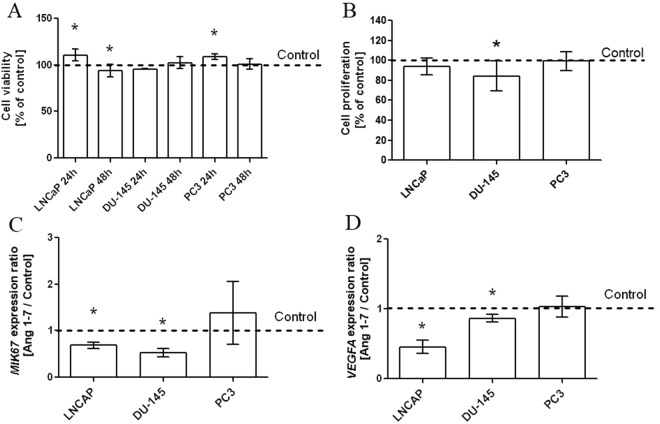
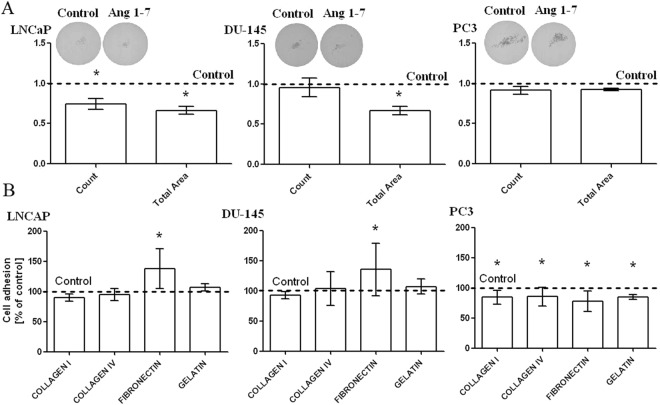
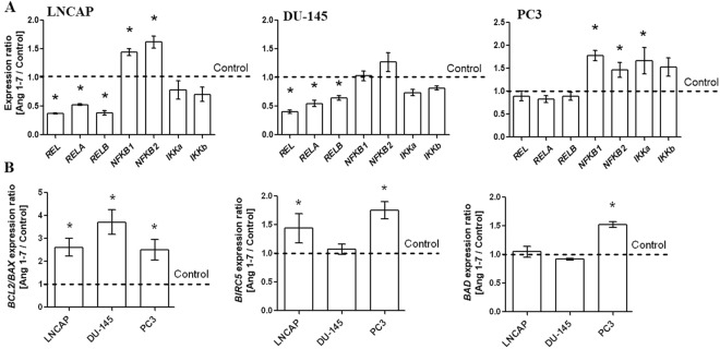
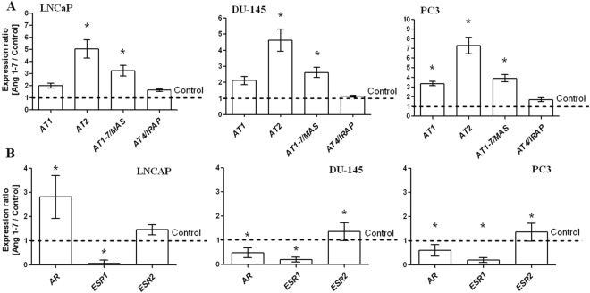
Angiotensin 1-7 (Ang1-7) is an endogenous bioactive component of the renin-angiotensin system (RAS). In addition to its cardiovascular properties, its anti-proliferative and anti-angiogenic traits are believed to play important roles in carcinogenesis. The present study examines the influence of Ang1-7 on processes associated with development and progression of prostate cancer cells. Our findings indicate that while Ang1-7 (1 nM; 48 h) can effectively reduce cell proliferation in DU-145, it can induce a significant decrease in the expression of MKI67 in LNCaP. In both cell lines we also observed a reduction in colony size in soft agar assay. A various changes in gene expression were noted after exposure to Ang1-7: those of anti- and pro-apoptotic agents and the NF-kB family of transcription factors, as well as mesenchymal cell markers and vascular endothelial growth factor A (VEGFA). In addition, Ang1-7 was found to modulate cell adhesion and matrix metallopeptidase (MMP) activity. Changes were also observed in the levels of angiotensin receptors and sex steroid hormone receptors. Ang1-7 reduced the levels of estrogen receptor alpha gene (ESR1) and increased the expression of estrogen receptor beta gene (ESR2) in all prostate cancer cells; it also up-regulated androgen receptor (AR) expression in androgen-sensitive cells but contradictory effect was observed in androgen- irresponsive cell lines. In summary, the results confirm the existence of complex network between the various elements of the local RAS and the molecular and cellular mechanisms of prostate cancerogenesis. The response of cancer cells to Ang1-7 appears to vary dependently on the dose and time of incubation as well as the aggressiveness and the hormonal status of cells.


Conflict of interest statement

The authors declare no competing interests.

Figures

Figure 1
Figure 1
Cell viability and proliferation of prostate cancer cells (LNCaP, DU-145, PC3) after exposure to Angiotensin 1–7 (mean ± SD; Tukey’s test: *p < 0.05). Panel (A) shows results of BrdU Assay and MTT Assay. Panel (B) presents results of expression of MKI67 and VEGFA genes.
Figure 2
Figure 2
The percentage of prostate cancer cells (LNCaP, DU-145, PC3) in the G0/G1, S, G2/M phases of cell cycle following exposure to Angiotensin 1–7 alone or in combination with angiotensin receptor inhibitors (I1: AT1 inhibitor - losartan; I2: AT2 inhibitor - PD123319; I3: AT1–7/MAS - A779; I4 – AT4/IRAP - HFI142) (mean ± SD; Tukey’s test: *p < 0.05).
Figure 3
Figure 3
The ability of adhesion and anchorage-independent cell growth after Ang1–7 treatment Panel (A) shows results of Soft Agar Colony Formation Assay (mean ± SEM; Tukey’s test: *p < 0.05). Panel (B) presents results of ECM Cell Adhesion Assay (mean ± SD; Tukey’s test: *p < 0.05).
Figure 4
Figure 4
Changes in the migration and invasion potential of the androgen-dependent (LNCaP) and androgen-independent prostate cancer cell lines (DU-145 and PC3) after exposure to Angiotensin 1–7. The left panel (A) presents the results of the wound healing assay. Panel (B) shows results of Gelatin Zymography to detect matrix metalloproteinase (MMP) activity The results in transwell migration/invasion assay are visible on the panel (C) (mean ± SD; Tukey’s test: *p < 0.05). Panel (D) present the expression of the EMT markers (mean ± SEM; Tukey’s test: *p < 0.05).
Figure 5
Figure 5
Expression of NFKB and IKK family genes (A) and expression of anti- and pro-apoptotic members (B) in the androgen-dependent (LNCaP) and androgen-independent prostate cancer cell lines (DU-145 and PC3) following exposure to Angiotensin 1–7 (mean ± SEM; Tukey’s test: *p < 0.05).
Figure 6
Figure 6
Expression of angiotensin receptor genes and expression of sex steroid hormone receptors in the androgen-dependent (LNCaP) and androgen-independent prostate cancer cell lines (DU-145 and PC3) following exposure to Angiotensin 1–7 (mean ± SEM; Tukey’s test: *p < 0.05).

Similar articles

Cited by

References

    1. Ohishi M, Yamamoto K, Rakugi H. Angiotensin (1–7) and other angiotensin peptides. Curr Pharm Des. 2013;19:3060–3064. doi: 10.2174/1381612811319170013. - DOI - PubMed
    1. Domińska K, Lachowicz-Ochedalska A. The involvement of the renin-angiotensin system (RAS) in cancerogenesis. Postepy Biochem. 2008;54:294–300. - PubMed
    1. Bernardi S, et al. Characterization and significance of ACE2 and Mas receptor in human colon adenocarcinoma. J Renin Angiotensin Aldosterone Syst. 2012;13:202–209. doi: 10.1177/1470320311426023. - DOI - PubMed
    1. Luo Y, et al. Expression of MAS1 in breast cancer. Cancer Sci. 2015;106:1240–1248. doi: 10.1111/cas.12719. - DOI - PMC - PubMed
    1. Ender SA, Dallmer A, Lässig F, Lendeckel U, Wolke C. Expression and function of the ACE2/angiotensin(1-7)/Mas axis in osteosarcoma cell lines U-2 OS and MNNG-HOS. Mol Med Rep. 2014;10:804–810. doi: 10.3892/mmr.2014.2266. - DOI - PubMed

Publication types

MeSH terms