. 2018 Oct 25;8(1):15787.
doi: 10.1038/s41598-018-34055-w.

In-vitro evaluation of apoptotic effect of OEO and thymol in 2D and 3D cell cultures and the study of their interaction mode with DNA

Affiliations

In-vitro evaluation of apoptotic effect of OEO and thymol in 2D and 3D cell cultures and the study of their interaction mode with DNA

Tahereh Jamali et al. Sci Rep. .

Abstract

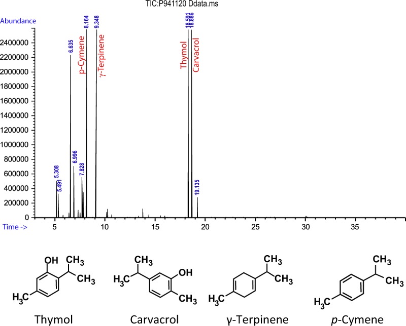
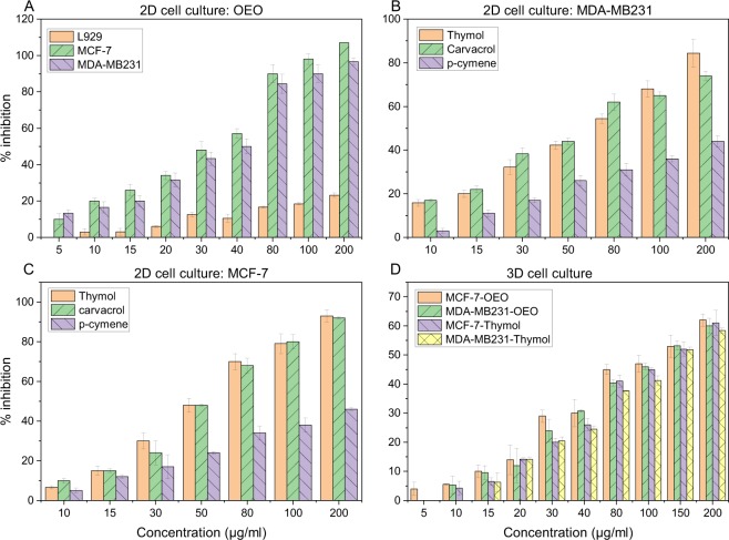
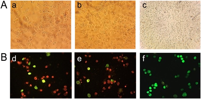
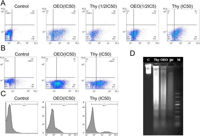
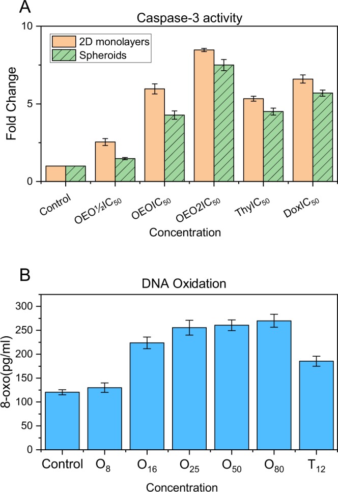
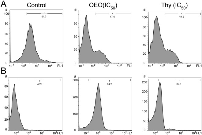
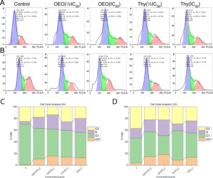
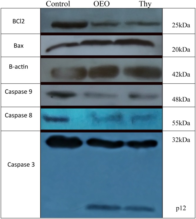
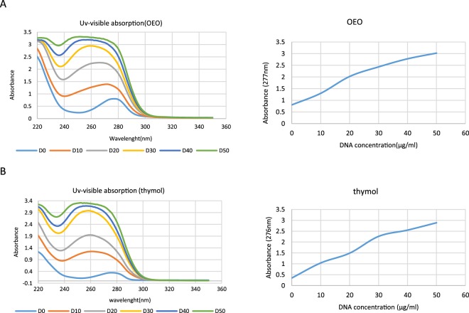
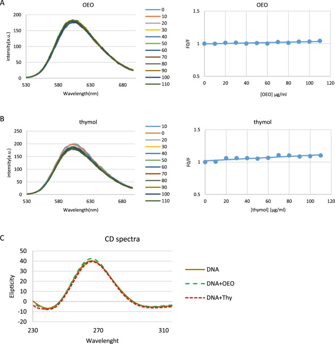
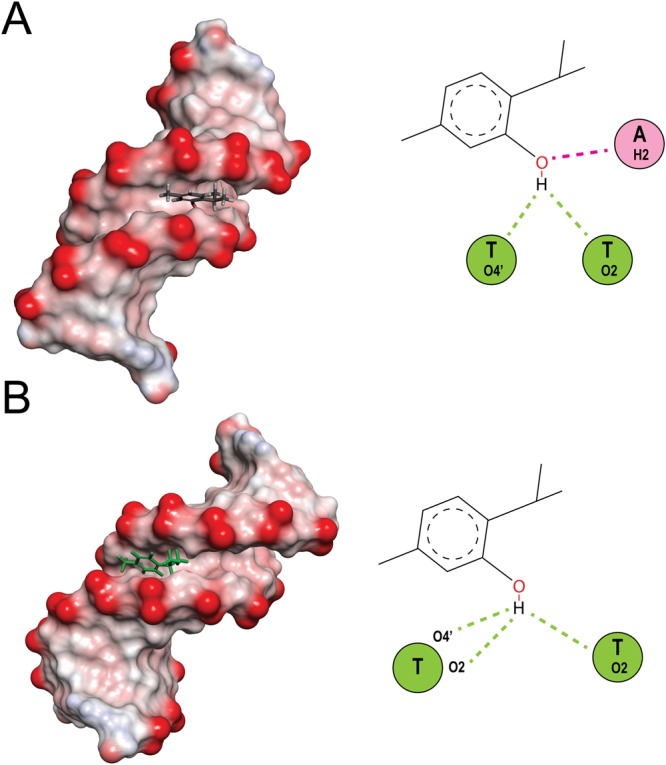
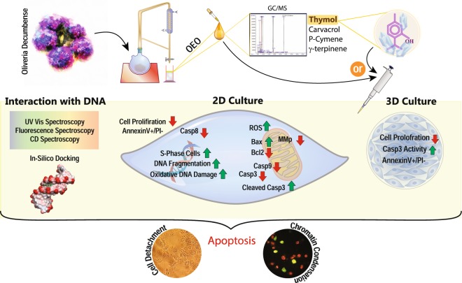
Oliveria decumbens is an Iranian endemic plant used extensively in traditional medicine. Recently, some studies have been performed on biological effects of Oliveria essential oil (OEO). However, to our knowledge, the anticancer activity of OEO has not been reported. Based on our GC/MS analysis, the basic ingredients of OEO are thymol, carvacrol, p-cymene and γ-terpinene. Therefore, we used OEO and its main component, thymol, to explore their effects on cell growth inhibition and anticancer activity. Despite having a limited effect on L929 normal cells, OEO/thymol induced cytotoxicity in MDA-MB231 breast cancer monolayers (2D) and to a lesser extent in MDA-MB231 spheroids (3D). Flow cytometry, caspase-3 activity assay in treated monolayers/spheroids and also fluorescence staining and DNA fragmentation in treated monolayers demonstrated apoptotic death mode. Indeed, OEO/thymol increased the Reactive Oxygen Species (ROS) level leading to mitochondrial membrane potential (MMP, ΔΨm) loss, caspase-3 activation and DNA damage caused S-phase cell cycle arrest. Furthermore, immunoblotting studies revealed the activation of intrinsic and maybe extrinsic apoptosis pathways by OEO/thymol. Additionally, in-vitro experiments, indicated that OEO/thymol interacts with DNA via minor grooves confirmed by docking method. Altogether, our reports underlined the potential of OEO to be considered as a new candidate for cancer therapy.


Conflict of interest statement

The authors declare no competing interests.

Figures

Figure 1
Figure 1
GC/MS chromatogram of OEO.
Figure 2
Figure 2
Spheroid formation in MDA-MB-231 (left) and MCF7 (right). they were imaged by inverted microscope.
Figure 3
Figure 3
Growth inhibition of (A) L929, MCF-7 and MDA-MB-231 monolayers after treatment with increasing concentrations of OEO for 24 h (B) MDA-MB-231 monolayers after treatment with increasing concentrations of thymol, carvacrol and p-cymene for 24 h and (C) MCF-7 monolayers after treatment with increasing concentrations of thymol, carvacrol and p-cymene for 24 h D) MDA-MB-231 and MCF-7 spheroids exposed to increasing concentrations of OEO and thymol for 24 h. Cell inhibition percentage and IC50 was determined using the MTT assay. The results are the means ± SDs from triplate experiments (P < 0.05).
Figure 4
Figure 4
Microscopic studies: (A) Morphological analysis of MDA-MB231 under inverted microscope. (a,b) the cells exposed to IC50 of OEO and thymol for 24 h; with morphological alterations including loss of adhesion, rounding, and sporadic distribution (c) control cells; with typical shape and a growth pattern of patchy monolayer. (B) Fluorescence staining of MDA-MB231 cells (d,e) the cells exposed to IC50 of OEO and thymol for 24 h (f) control cells. The cells were stained using AO and EtBr and visualized with fluorescence microscopy.
Figure 5
Figure 5
Annexin-V/PI analysis by flow cytometry in (A) MDA-MB231 monolayer cells (2D): control and treated cells with OEO (IC50 and 1/2IC50), thymol (IC50 and 1/2IC50). (B) MDA-MB231 cells separated from spheroids (3D): control and treated cells with OEO (IC50) and thymol (IC50.). (C) DNA fragmentation in treated MDA-MB-231: TUNEL assay, apoptotic intensity of MDA-MB231 cells was determined by flow cytometry after TUNEL assay. (A) Shift of the population to the right in treated cells compared to control cells indicates the apoptotic cell population. (D) DNA fragmentation in treated MDA-MB-231: DNA laddering, Lane 1: Control, Lane 2: the cells treated with thymol (IC50); Lane 3: the cells treated with OEO (IC50). Lane 4: the cells treated with positive control (IC50) and Lane 5: molecular marker.
Figure 6
Figure 6
(A) Caspase 3 activity assay in control and treated cells with OEO (1/2IC50, IC50 and 2IC50), thymol (IC50) and Doxorubicin (IC50) in A: MDA-MB231 monolayer culture (2D) and MDA-MB231 spheroids (3D). The data were expressed in fold change with respect to untreated control groups. (B) Levels of 8-oxo-dG in the control and treated MDA-MB231 cells with various concentrations of OEO and thymol.
Figure 7
Figure 7
(A) Effect of OEO and thymol on Δψm in MDA-MB-231 control cells and treated cells with IC50 of OEO and thymol. The histograms reveal a left shift of peak demonstrated the decrease of Rh-123 fluorescence intensity because of the loss of MMP. (B) Effect of OEO and thymol on the formation of reactive oxygen species (ROS) in MDA-MB-231 control cells and treated cells with IC50 of OEO and thymol respectively. The histograms with a right shift reveal the increase of ROS level in treated MDA-MB231 cells with OEO and thymol.
Figure 8
Figure 8
(A,B) Effect of OEO and thymol on DNA content of MDA-MB-231 cells after 4 h (top) and 12 h (bottom). (C,D) Measurement of cell cycle populations after treatment with OEO and thymol after 4 h and 12 h. Cell cycle analysis are done using a DNA intercalating dye, such as PI, to quantify the amount of DNA present in each cell.
Figure 9
Figure 9
OEO/thymol-mediated apoptosis of MDA-MB231 cells involves a caspase-dependent mechanism and mitochondrial stress. MDA-MB231 cells were exposed to the IC50 of the OEO and thymol, and the expression of procaspases, Bax and Bcl2 were revealed by Western blot analysis.
Figure 10
Figure 10
Interaction of (A) OEO and (B) thymol with dsDNA using UV–visible spectroscopy. UV–visible absorption spectra of OEO and thymol (50 µg/ml) in presence of increasing concentrations of dsDNA (0–50 µg/ml) in phosphate buffer (0.1 M with pH = 7.4).
Figure 11
Figure 11
(A and B) Competitive displacement assays. Fluorescence titration of EtBr–dsDNA complex with increasing concentrations of (A) OEO and (B) thymol. No significant effect of OEO and thymol was seen on EtBr-dsDNA system. Right plots are Stern–Volmer plots for the mechanism of fluorescence quenching of EtBr–DNA by OEO and thymol. (C) Effect of OEO and thymol on CD spectra of dsDNA. CD spectra of dsDNA (50 µg/ml in phosphate buffer (0.1 M with pH = 7.4)) in presence of IC50 of OEO and thymol.
Figure 12
Figure 12
Molecular docked models of (A) thymol and (B) carvacrol with DNA (PDB 1D: 1BNA). Conventional hydrogen bonds and carbon hydrogen bonds have been labeled using green and pink dashed lines respectively.
Figure 13
Figure 13
Schematic representation of OEO/thymol-induced apoptosis.

References

    1. Enayatrad M, Amoori N, Salehiniya H. Epidemiology and trends in breast cancer mortality in Iran. Iranian journal of public health. 2015;44:430. - PMC - PubMed
    1. Maruthanila V, Elancheran R, Kunnumakkara A, Kabilan S, Kotoky J. Recent development of targeted approaches for the treatment of breast cancer. Breast Cancer. 2017;24:191. doi: 10.1007/s12282-016-0732-1. - DOI - PubMed
    1. Emami SA, et al. Growth Inhibition and Apoptosis Induction of Essential Oils and Extracts of Nepeta cataria L on Human Prostatic and Breast Cancer Cell Lines. Asian Pacific journal of cancer prevention. 2016;17:125. doi: 10.7314/APJCP.2016.17.S3.125. - DOI - PubMed
    1. Lahlou M. The success of natural products in drug discovery. Pharmacol Pharm. 2013;4:17. doi: 10.4236/pp.2013.43A003. - DOI
    1. Roy A, Attre T, Bharadvaja N. Anticancer agent from medicinal plants: a review. New apects in medicinal plants and pharmacognosy. 2017;1:54.

MeSH terms