. 2019 Feb 18;213(0):453-469.
doi: 10.1039/c8fd00097b.

Computing of temporal information in spiking neural networks with ReRAM synapses

Affiliations

Computing of temporal information in spiking neural networks with ReRAM synapses

W Wang et al. Faraday Discuss. .

Abstract

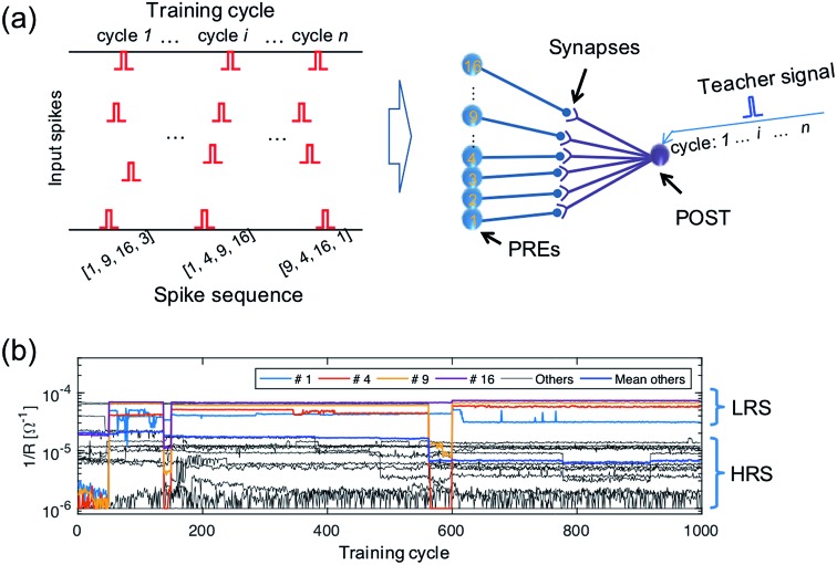
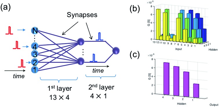
Resistive switching random-access memory (ReRAM) is a two-terminal device based on ion migration to induce resistance switching between a high resistance state (HRS) and a low resistance state (LRS). ReRAM is considered one of the most promising technologies for artificial synapses in brain-inspired neuromorphic computing systems. However, there is still a lack of general understanding about how to develop such a gestalt system to imitate and compete with the brain's functionality and efficiency. Spiking neural networks (SNNs) are well suited to describe the complex spatiotemporal processing inside the brain, where the energy efficiency of computation mostly relies on the spike carrying information about both space (which neuron fires) and time (when a neuron fires). This work addresses the methodology and implementation of a neuromorphic SNN system to compute the temporal information among neural spikes using ReRAM synapses capable of spike-timing dependent plasticity (STDP). The learning and recognition of spatiotemporal spike sequences are experimentally demonstrated. Our simulation study shows that it is possible to construct a multi-layer spatiotemporal computing network. Spatiotemporal computing also enables learning and detection of the trace of moving objects and mimicking of the hierarchy structure of the biological visual cortex adopting temporal-coding for fast recognition.


Figures

Fig. 1
Fig. 1. (a) Illustration of a building block of the biological neural system, consisting of a pre-synaptic neuron (PRE), a post-synaptic neuron (POST), and a synapse between the PRE axon and POST dendrite. (b) Comparison between the biological synapse (left) and electronic synapse (right, resistive switching memory, ReRAM).
Fig. 2
Fig. 2. (a) Schematic view of the temporal computation between the two spikes using ReRAM synapses. To enable the interference of the two temporally separated PRE spikes, the PREs convert the spike signals into exponentially decaying voltage signals applied to the gate of the one-transistor/one-ReRAM (1T1R) synapses. Inset: the conductance of the two ReRAM devices. (b) The exponentially decaying signals of the output of the two PREs (upper panel) and the internal potential (Vint) of the POST (lower panel). (c) Maximum Vint as a function of the temporal correlation (the time difference of the two PRE spikes, tc).
Fig. 3
Fig. 3. (a) The building block in a fully functional neuromorphic system implementing the weight update algorithm. Inset: The IV characteristics of a single ReRAM device. (b–d) The weight updating rule implemented in the neuromorphic building block, showing: (b) the PRE signal applied to the gate terminal of 1T1R synapse; (c) the voltage applied to the top electrode of the ReRAM device, generated by the supervisor circuit; and (d) the weight update of the synapse. The direction of the weight update is decided by the polarity of the top electrode voltage, and the amount of weight change is related to the gate voltage of the transistor at the time of updating, which incorporates the temporal information of the PRE spikes.
Fig. 4
Fig. 4. (a) A schematic illustration of the input spiking patterns submitted to a 16 × 1 spatiotemporal network supervised by a teacher signal. (b) The experimentally measured evolution of the synaptic weights during training.
Fig. 5
Fig. 5. Measured Vint, indicating spike accumulation by the POST for (a) the true sequence and (b) false sequences.
Fig. 6
Fig. 6. (a) Illustration of a two-layer spatiotemporal network to recognize a relatively long spiking sequence. (b and c) The conductance map of the synapses in the two-layer network.
Fig. 7
Fig. 7. (a and b) The internal potential of the hidden layer neurons (a) and of the output neuron (b) under the submission of the true sequence. (c and d) The same, but under the submission of a false sequence.
Fig. 8
Fig. 8. (a) Illustration of a neural network for the mapping of a spatiotemporal pattern. (b) Randomly generated spatiotemporal input pattern; (c) spatiotemporal output pattern; (d) the training of mapping a complex spatiotemporal pattern to a simple one.
Fig. 9
Fig. 9. (a) Illustration of a dynamic “X” pattern moving horizontally (first row) or diagonally (second row). (b) Illustration of the two-layer neural network for the recognition of a moving object.
Fig. 10
Fig. 10. (a) Illustration of the hierarchy structure of the biological visual system. (b) Schematic diagram of the artificial visual system for feature extraction and classification using spatiotemporal spike coding.
Fig. 11
Fig. 11. (a) The optical digital character for training (clean pattern) and testing (noise pattern) of the artificial hierarchical visual neural network. (b) The recognition rate of the trained network as a function of the noise level of the optical digital character.

References

    1. LeCun Y., Bengio Y., Hinton G. Nature. 2015;521:436–444. - PubMed
    1. Shulaker M. M., Hills G., Park R. S., Howe R. T., Saraswat K., Wong P., Mitra S. Nature. 2017;547:74–78. - PubMed
    1. Esser S. K., Merolla P. A., Arthur J. V., Cassidy A. S., Appuswamy R., Andreopoulos A., Berg D. J., McKinstry J. L., Melano T., Barch D. R., di Nolfo C., Datta P., Amir A., Taba B., Flickner M. D., Modha D. S. Proc. Natl. Acad. Sci. U. S. A. 2016;113:11441–11446. - PMC - PubMed
    1. Merolla P. A., V Arthur J., Alvarez-icaza R., Cassidy A. S., Sawada J., Akopyan F., Jackson B. L., Imam N., Guo C., Nakamura Y., Brezzo B., Vo I., Esser S. K., Appuswamy R., Taba B., Amir A., Flickner M. D., Risk W. P., Manohar R., Modha D. S. Science. 2014;345:668–673. - PubMed
    1. Yang J. J., Strukov D. B., Stewart D. R. Nat. Nanotechnol. 2013;8:13–24. - PubMed

Publication types