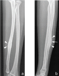
Post-traumatic osteoid osteoma in an 18-year-old adolescent
- PMID: 30363164
- PMCID: PMC6159126
- DOI: 10.1259/bjrcr.20150141
Post-traumatic osteoid osteoma in an 18-year-old adolescent
Abstract
Osteoid osteoma (OO) is a painful, benign bone-forming lesion, which often poses a diagnostic challenge. The aetiology of OO is still poorly understood. Although not generally accepted, an association with previous trauma or infection has occasionally been suggested. We present a case of an OO 12 years following an ulnar fracture. Radiologists should consider OO as a potential delayed "complication" of a previous fracture. Persistent pain at a previous fracture site should alert the clinician to request cross-sectional imaging. CT scanning plays a pivotal role in the correct diagnosis of OO.
Figures


References
-
- Chai JW, Hong SH, Choi JY, Koh YH, Lee JW, Choi JA, et al. . . Radiologic diagnosis of osteoid osteoma: from simple to challenging findings. Radiographics 2010; 30: 737–49. - PubMed
-
- Baron D, Soulier C, Kermabon C, Leroy JP, Le Goff P. . [Post-traumatic osteoid osteoma. apropos of 2 cases and review of the literature]. Rev Rhum Mal Osteoartic 1992; 59: 271–5. - PubMed
-
- Adil A, Hoeffel C, Fikry T. . Osteoid osteoma after a fracture of the distal radius. Am J Roentgenol 1996; : 145–6. - PubMed
-
- Soon M, Low CK, Chew J. . Osteoid osteoma after a stress fracture of the tibia: a case report. Ann Acad Med Singapore 1999; : 877–8. - PubMed
-
- Grenard N, Duparc F, Roussignol X, Chomant J, Muller JM, Dujardin F, et al. . . Osteoid osteoma after an old femoral shaft fracture. Rev Chir Orthop Reparatrice Appar Mot 2001; : 503–5. - PubMed
Publication types
LinkOut - more resources
Full Text Sources
Other Literature Sources

