

Case Reports
doi: 10.1259/bjrcr.20170029.
eCollection 2018.
Atypical hepatic haemangiomas
Affiliations
- PMID: 30363183
- PMCID: PMC6159142
- DOI: 10.1259/bjrcr.20170029
Item in Clipboard
Case Reports
Atypical hepatic haemangiomas
BJR Case Rep.
.
Abstract
Hepatic haemangioma is the most common benign liver lesion in the general population. It often exhibits a uniform pattern of characteristics, thus being called "typical." However, a certain number of hepatic haemangiomas have special or uncommon characteristics and are termed "atypical." The majority of patients are asymptomatic. Its differential diagnosis is critical, and its differentiation from other aetiological possibilities can be challenging, especially in cases of atypical haemangiomas, which may lead to confusion or even misleading diagnoses. We report on a 55-year-old patient with atypical multiple hepatic haemangiomas mimicking metastasis or echinococcus infection.
Figures
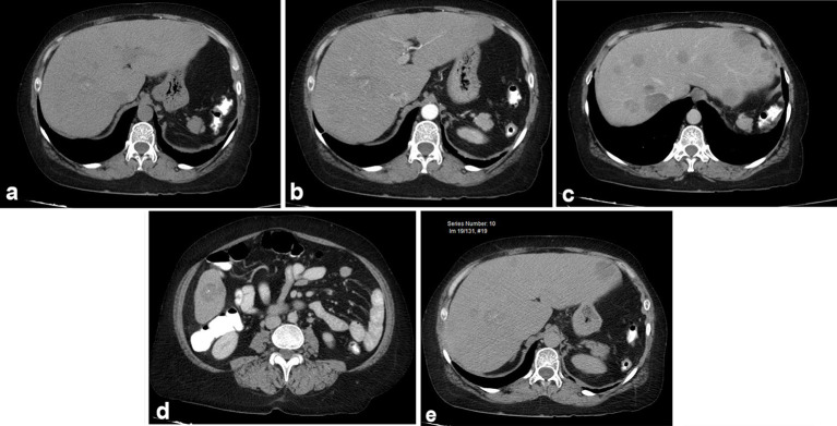
Abdominal CT showing multiple hepatic lesions ranging from a few to 5 cm, which do not significantly enhance and contain microcalcifications. (a) Simple phase: focal lesions are hypodense with microcalcifications (arrow); (b) arterial phase, without significant enhancement: no hypervascular lesions are identified. (c and d) Portal phase: discrete uptake; (e) late phase: persisting hypodense lesions without retention of contrast medium.
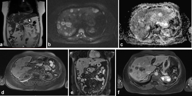
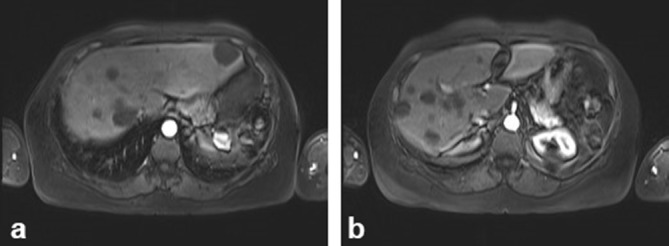
MRI showing a similar pattern of enhancement. (a) Coronal sequence, T2 Half Fourier Single Shot Turbo-spin Echo, (HASTE): normal-sized liver with multiple, hyperintense lesions in all liver segments; (b and c) diffusion sequences: lesions show restriction predominantly in the periphery; (d) T1 fat suppression without contrast media: hypointense lesions in the liver; (e) T1 coronal sequence in portal phase: the lesions remain without significant uptake of contrast medium; (f) Axial T1 late phase, 5 min after administration of contrast medium: the lesions with some small zones of central uptake.
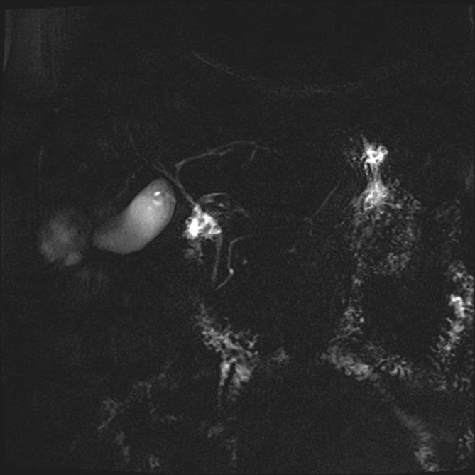
Cholangioresonance sequences.
(a) MRI axial T1 late phase, 5 min after administration of contrast medium, showing low uptake of the contrast medium, which is heterogeneous; central sinus is seen in some lesions and peripheral in others. (b) MRI axial T1 late phase, 5 min. Lesions show little contrast medium uptake, simulating lesions of a cystic nature.
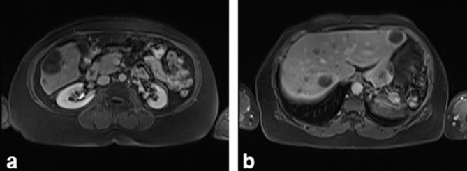
(a, b) MRI axial, arterial phase: multiple low-intensity lesions are identified that do not show contrast medium uptake.
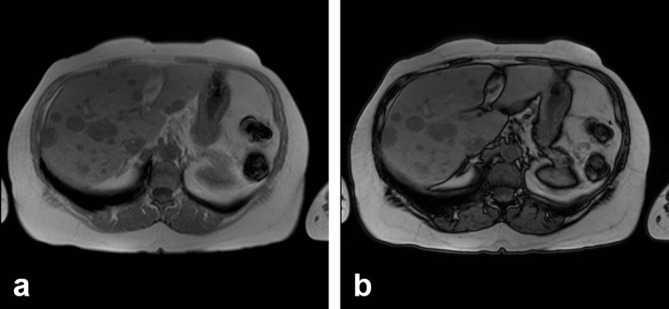
(a and b) Phase and out-of-phase sequences were performed without identifying change in signal intensity, suggesting microscopic fat content in the lesions.
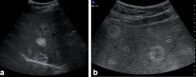
Liver Ultrasound: (a and b) Multiple heterogeneous, predominantly echogenic hepatic lesions were identified, distributed among all liver segments. Some had target-like or annular appearance and others had calcifications. Some had a pattern similar to “hailstorm” described in echinococcosis.
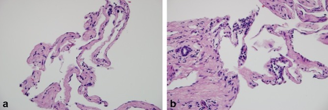
Liver biopsy. (a) Vascular channels lined by endothelial cells without dysplasia, separated by septa of fibrous enlargement (100×); (b) Vascular channels adjacent to a portal space that presents occasional lymphocytes and fibrosis (200×).
References
-
- Vilgrain V, Boulos L, Vullierme MP, Denys A, Terris B, Menu Y. Imaging of atypical hemangiomas of the liver with pathologic correlation. Radiographics 2000; 20: 379–97. - PubMed
-
- Curry MP, Chopra S.. Hepatic hemangioma. Uptodate 2015; 1–18.
-
- Klotz T, Montoriol PF, Da Ines D, Petitcolin V, Joubert-Zakeyh J, Garcier JM. Hepatic haemangioma: common and uncommon imaging features. Diagn Interv Imaging 2013; 94: 849–59. - PubMed
-
- Sánchez T FA, Zugbe G N, Lúcia C ME, Moraga L M.. Hemangioma hepático poliquístico simulando un quiste hidatídico: Reporte de un caso y revisión del tema. Revista chilena de radiología 2014; 20: 164–7.
-
- Dres Jose Palau AB, Belaunzarán A, Saiz EG.. Hallazgos en imagen del hemangioma hepático. Revista del Hospital Privado de Comunidad 2006; 9: 19–23.
Publication types
LinkOut - more resources
Full Text Sources
