

Case Reports
doi: 10.1259/bjrcr.20160094.
eCollection 2017.
Zinner syndrome: an unusual cause of bladder outflow obstruction
Affiliations
- PMID: 30363237
- PMCID: PMC6159234
- DOI: 10.1259/bjrcr.20160094
Item in Clipboard
Case Reports
Zinner syndrome: an unusual cause of bladder outflow obstruction
BJR Case Rep.
.
Abstract
Zinner syndrome is a rare condition comprising a triad of unilateral renal agenesis, ipsilateral seminal vesicle obstruction and ipsilateral ejaculatory duct obstruction. The mutual embryological origins of the seminal vesicle and the ureteral bud result in both anomalous genital and urinary tracts. We present the case of a 39-year-old patient where the initial presentation of this condition was bladder outflow obstruction. In this paper, we discuss the embryological origin of this condition, the range of imaging tools used to diagnose Zinner syndrome and the inherent benefits and shortcomings of each modality.
Figures
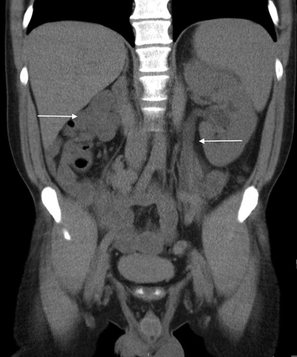
CT coronal view showing hypoplastic right kidney and dilated left ureter and mild hydronephrosis.

Axial T2 weighted MRI image showing thickened bladder and dilated, cystic right ureteric bud.

Coronal T2 weighted MRI image showing thickened bladder (B), cystic right ureteric bud opening in the bladder (arrow) and dilated seminal vesicle (S).

Coronal T2 weighted MRI image showing seminal vesicle cyst causing occlusion of bladder neck.

Axial T2 weighted MRI image showing dilated, tortuous seminal vesicle.

Sagittal T2 weighted MRI image showing dilated left ureter.
Similar articles
-
Zinner syndrome presenting with intermittent scrotal pain in a young man.Radiol Case Rep. 2018 Sep 20;13(6):1224-1227. doi: 10.1016/j.radcr.2018.08.012. eCollection 2018 Dec. Radiol Case Rep. 2018. PMID: 30258511 Free PMC article.
-
Zinner Syndrome.Eur J Case Rep Intern Med. 2021 Jun 3;8(7):002628. doi: 10.12890/2021_002628. eCollection 2021. Eur J Case Rep Intern Med. 2021. PMID: 34268266 Free PMC article.
-
A rare case of Zinner syndrome: Triad of unilateral renal agenesis, ipsilateral seminal vesicle cyst and ejaculatory duct obstruction.Radiol Case Rep. 2021 Aug 30;16(11):3380-3382. doi: 10.1016/j.radcr.2021.08.012. eCollection 2021 Nov. Radiol Case Rep. 2021. PMID: 34504629 Free PMC article.
-
Zinner syndrome: two cases and review of the literature.BMJ Case Rep. 2021 Jun 17;14(6):e243002. doi: 10.1136/bcr-2021-243002. BMJ Case Rep. 2021. PMID: 34140330 Free PMC article. Review.
-
Classifying seminal vesicle cysts in the diagnosis and treatment of Zinner syndrome: A report of six cases and review of available literature.Andrologia. 2020 Feb;52(1):e13397. doi: 10.1111/and.13397. Epub 2019 Nov 15. Andrologia. 2020. PMID: 31729082 Review.
Cited by
-
Zinner syndrome unveiled: Ectopic ureter and seminal vesicle cyst leading to urinary dysfunction: A case report.Radiol Case Rep. 2025 Jan 7;20(3):1721-1725. doi: 10.1016/j.radcr.2024.12.017. eCollection 2025 Mar. Radiol Case Rep. 2025. PMID: 39868060 Free PMC article.
-
Incidental imaging findings suggesting Zinner syndrome in a young patient with pulmonary embolism: A case report.Radiol Case Rep. 2020 Feb 19;15(4):437-441. doi: 10.1016/j.radcr.2020.01.027. eCollection 2020 Apr. Radiol Case Rep. 2020. PMID: 32148603 Free PMC article.
-
Zinner Syndrome: A Case Report of Rare Urogenital Anomaly.J Med Ultrasound. 2021 May 4;30(1):59-61. doi: 10.4103/JMU.JMU_125_20. eCollection 2022 Jan-Mar. J Med Ultrasound. 2021. PMID: 35465594 Free PMC article.
-
Zinner syndrome mimicking bladder outlet obstruction managed with aspiration.Urol Ann. 2019 Oct-Dec;11(4):449-452. doi: 10.4103/UA.UA_152_18. Urol Ann. 2019. PMID: 31649472 Free PMC article.
-
Zinner syndrome in adult male: a rare case report.Transl Androl Urol. 2025 Mar 30;14(3):848-854. doi: 10.21037/tau-2024-763. Epub 2025 Mar 26. Transl Androl Urol. 2025. PMID: 40226079 Free PMC article.
References
-
- Zinner A. Ein fall von intravesikaler Samenblasenzyste. Wien Med Wochenschr. 1914; 64: 605.
-
- Moore KL. The developing human, 2nd ed Philadelphia: Saunders, 1997:220-246.
-
- Williams RD, Sandlow JI.. Surgery of the seminal vesicles : Walsh PC, Retik AB Jr Vaughan ED, Wein AJ, . Campbell's urology. 7th ed Philadelphia: Saunders; 1998. 3299–307.
-
- Livingston L, Larsen CR.. Seminal vesicle cyst with ipsilateral renal agenesis. AJR Am J Roentgenol 2000; 175: 177–80. - PubMed
Publication types
LinkOut - more resources
Full Text Sources
