

Prostate imaging features that indicate benign or malignant pathology on biopsy
- PMID: 30363462
- PMCID: PMC6178322
- DOI: 10.21037/tau.2018.07.06
Prostate imaging features that indicate benign or malignant pathology on biopsy
Abstract
Accurate diagnosis of clinically significant prostate cancer is essential in identifying patients who should be offered treatment with curative intent. Modifications to the Gleason grading system in recent years show that accurate grading and reporting at needle biopsy can improve identification of clinically significant prostate cancers. Extracapsular extension of prostate cancer has been demonstrated to be an adverse prognostic factor with greater risk of metastatic spread than organ-confined disease. Tumor volume may be an independent prognostic factor and should be considered in conjunction with other factors. Multi-parametric magnetic resonance imaging (MP-MRI) has become an increasingly important tool in the diagnosis and characterization of prostate cancer. MP-MRI allows T2-weighted (T2W) anatomical imaging to be combined with functional and physiological assessment. Diffusion-weighted imaging (DWI) has shown greater sensitivity, specificity and negative predictive value compared to prostate specific antigen (PSA) testing and T2W imaging alone and has a more positive correlation with Gleason score and tumour volume. Dynamic gadolinium contrast-enhanced (DCE) imaging can exhibit difficulties in distinguishing prostatitis from malignancy in the peripheral zone, and between benign prostatic hyperplasia (BPH) and malignancies in the transition zone (TZ). Computer aided diagnosis utilizes software to aid radiologists in detecting and diagnosing abnormalities from diagnostic imaging. New techniques of quantitative MRI, such as VERDICT MRI use tissue-specific factors to delineate different cellular and microstructural phenotypes, characterizing tissue properties with greater detail. Proton MR spectroscopic imaging (MRSI) is a more technically challenging imaging modality than DCE and DWI MRI. Over the last decade, choline and prostate-specific membrane antigen (PSMA) positron emission tomography (PET) have developed as better tools for staging than conventional imaging. While hyperpolarized MRI shows promise in improving the imaging and differentiation of benign and malignant lesions there is further work required. Accurate reading and interpretation of diagnostic investigations is key to accurate identification of abnormal areas requiring biopsy, sparing those in whom benign or indolent disease can be managed by non-invasive means. Embracing and advancing existing technologies is essential in furthering this process.
Keywords: Prostate; benign; imaging; magnetic resonance imaging (MRI); malignant; parametric; qualitative; quantitative.
Conflict of interest statement
Conflicts of Interest: HU Ahmed proctors for HIFU and cryotherapy and is paid for training other surgeons in these procedures. HU Ahmed is a paid medical consultant for Sophiris Biocorp and Sonacare Inc. M Winkler receives a travel grant and a loan of device from Zicom Biobot. Other authors have no conflicts of interest to declare.
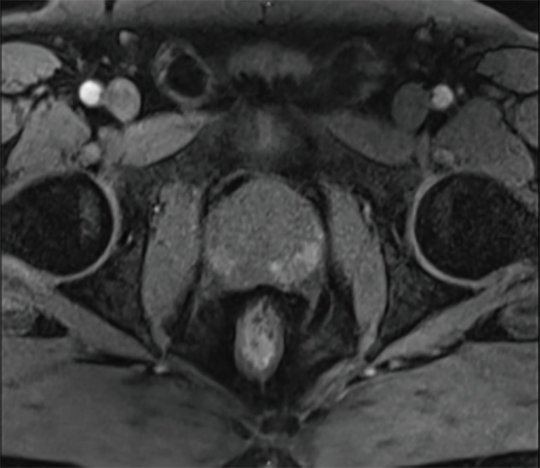
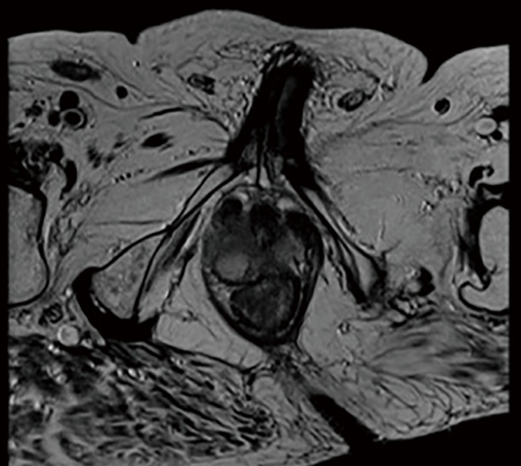
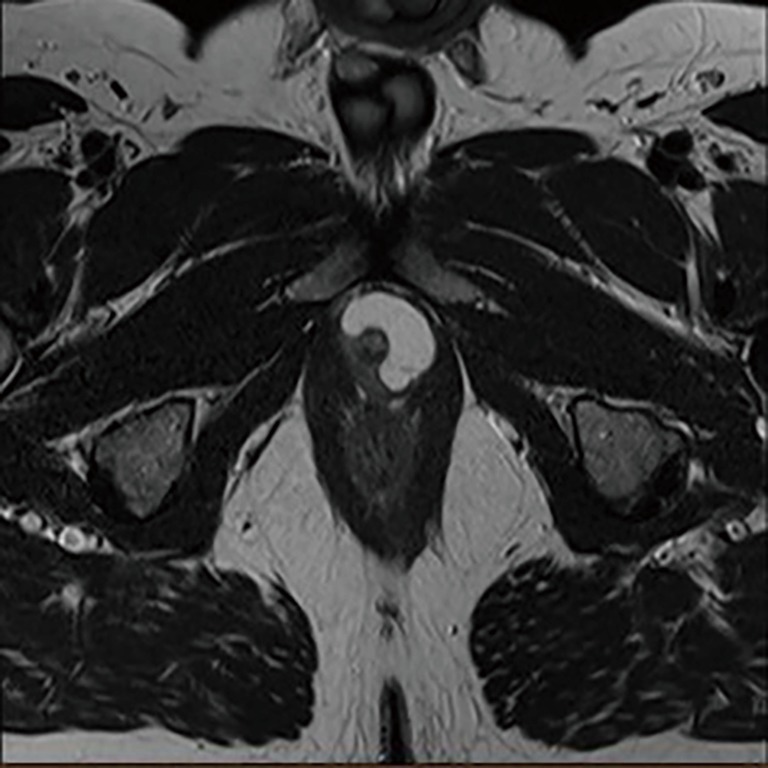
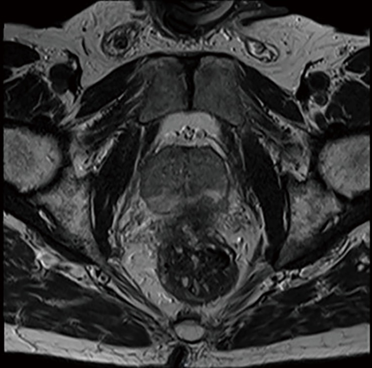
Figures
Similar articles
-
More advantages in detecting bone and soft tissue metastases from prostate cancer using 18F-PSMA PET/CT.Hell J Nucl Med. 2019 Jan-Apr;22(1):6-9. doi: 10.1967/s002449910952. Epub 2019 Mar 7. Hell J Nucl Med. 2019. PMID: 30843003
-
Multiparametric Magnetic Resonance Imaging in Evaluation of Clinically Significant Prostate Cancer.Indian J Radiol Imaging. 2021 Jan;31(1):65-77. doi: 10.1055/s-0041-1730093. Epub 2021 May 23. Indian J Radiol Imaging. 2021. PMID: 34316113 Free PMC article.
-
Multiparametric MRI in detection and staging of prostate cancer.Dan Med J. 2017 Feb;64(2):B5327. Dan Med J. 2017. PMID: 28157066 Review.
-
Development and validation of a logistic regression model to distinguish transition zone cancers from benign prostatic hyperplasia on multi-parametric prostate MRI.Eur Radiol. 2017 Sep;27(9):3600-3608. doi: 10.1007/s00330-017-4775-2. Epub 2017 Mar 13. Eur Radiol. 2017. PMID: 28289941
-
Clinical and imaging tools in the early diagnosis of prostate cancer, a review.JBR-BTR. 2010 Mar-Apr;93(2):62-70. doi: 10.5334/jbr-btr.121. JBR-BTR. 2010. PMID: 20524513 Review.
Cited by
-
Detection of High-Grade Prostate Cancer With a Super High B-value (4000 s/mm2) in Diffusion-Weighted Imaging Sequences by Magnetic Resonance Imaging.Cureus. 2022 Mar 3;14(3):e22807. doi: 10.7759/cureus.22807. eCollection 2022 Mar. Cureus. 2022. PMID: 35399424 Free PMC article.
-
Primary Neuroendocrine Tumor of Prostate in a Case of Metastatic Adenocarcinoma of Lung: Rare Entity with Histopathological and Gallium 68 DOTANOC Positron Emission Tomography Correlation.Indian J Nucl Med. 2023 Apr-Jun;38(2):154-156. doi: 10.4103/ijnm.ijnm_193_22. Epub 2023 Jun 8. Indian J Nucl Med. 2023. PMID: 37456183 Free PMC article.
-
Parametric maps of spatial two-tissue compartment model for prostate dynamic contrast enhanced MRI - comparison with the standard tofts model in the diagnosis of prostate cancer.Phys Eng Sci Med. 2023 Sep;46(3):1215-1226. doi: 10.1007/s13246-023-01289-6. Epub 2023 Jul 11. Phys Eng Sci Med. 2023. PMID: 37432557
-
Meta Analysis of Efficacy and Safety of Prostate Biopsy: A Comparison Between Transperineal and Transrectal Approach.Urol Res Pract. 2025 Jan 3;50(4):208-218. doi: 10.5152/tud.2025.24094. Urol Res Pract. 2025. PMID: 39873401 Free PMC article. Review.
-
Parametric maps of spatial two-tissue compartment model for prostate dynamic contrast enhanced MRI - comparison with the standard Tofts model in the diagnosis of prostate cancer.Res Sq [Preprint]. 2023 Feb 8:rs.3.rs-2539644. doi: 10.21203/rs.3.rs-2539644/v1. Res Sq. 2023. Update in: Phys Eng Sci Med. 2023 Sep;46(3):1215-1226. doi: 10.1007/s13246-023-01289-6. PMID: 36798227 Free PMC article. Updated. Preprint.
References
Publication types
Grants and funding
LinkOut - more resources
Full Text Sources
Research Materials
Miscellaneous