Review
. 2018 Sep;7(Suppl 4):S462-S476.
doi: 10.21037/tau.2018.06.09.

Role of PET imaging for biochemical recurrence following primary treatment for prostate cancer

Affiliations
Review

Role of PET imaging for biochemical recurrence following primary treatment for prostate cancer

Samuel J Galgano et al. Transl Androl Urol. 2018 Sep.

Abstract

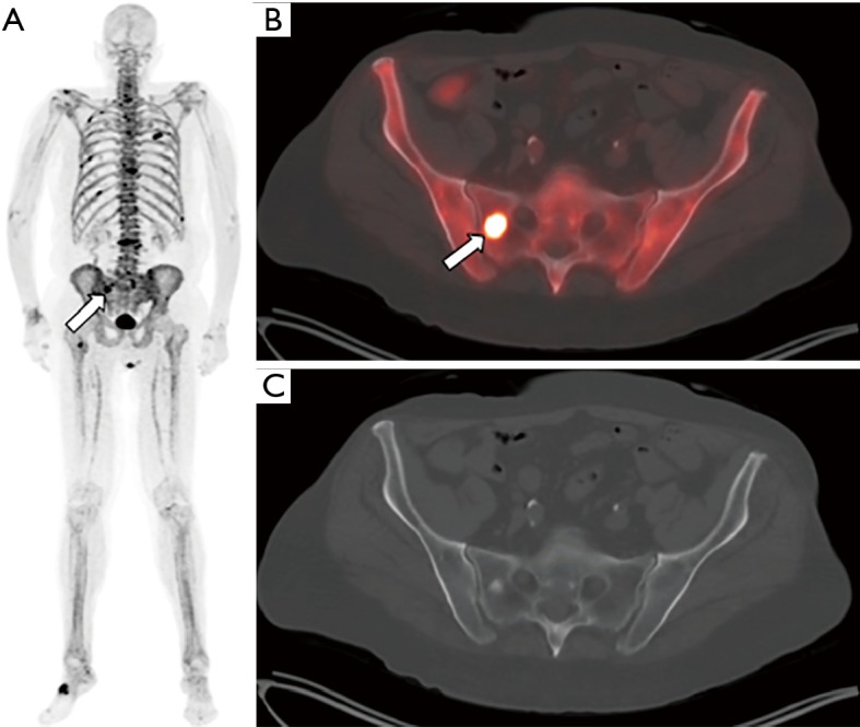
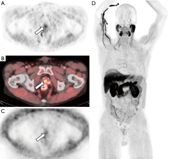
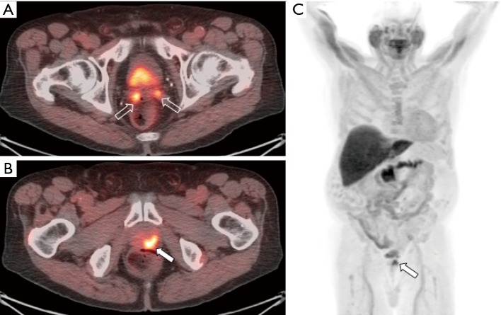
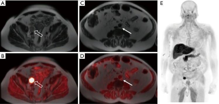
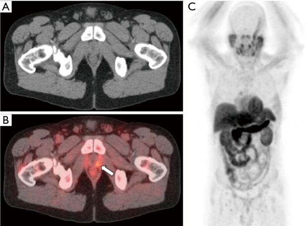
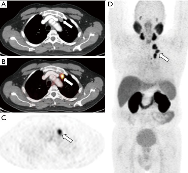
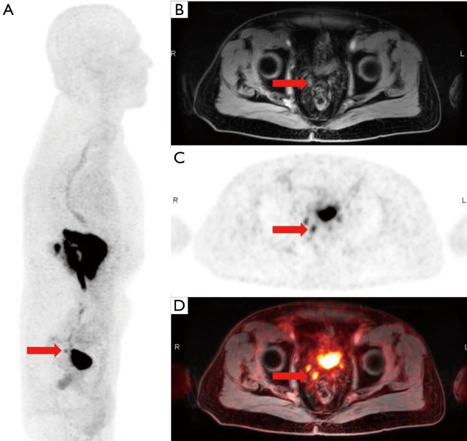
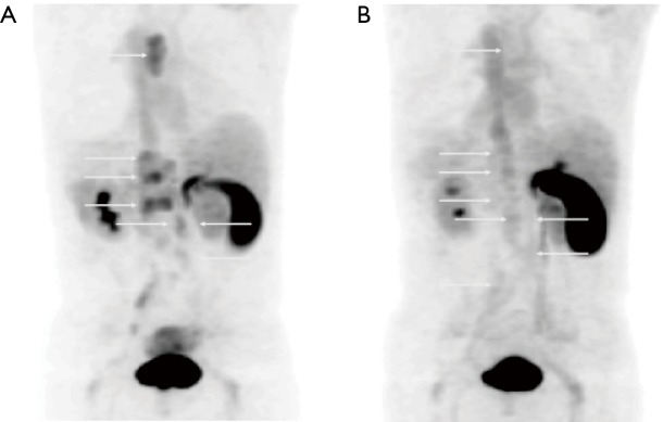
Prostate cancer is one of the most common cancers in men worldwide, and primary prostate cancer is typically treated with surgery, radiation, androgen deprivation, or a combination of these therapeutic modalities. Despite technical advances, approximately 30% of men will experience biochemical recurrent within 10 years of definitive treatment. Upon detection of a rise in serum prostate specific antigen (PSA), there is great need to accurately stage these patients to help guide further therapy. As a result, there are considerable efforts underway to establish the role of positron emission tomography (PET) in the diagnostic algorithm of biochemically recurrent prostate cancer. This manuscript provides an overview of PET tracers used for the detection and localization of prostate cancer in the setting of biochemical recurrence with a focus on PET tracers that are currently being used in clinical practice in the United States.

Keywords: Biochemical recurrence; PET/magnetic resonance imaging (MRI); positron emission tomography (PET)/computed tomography (CT); prostate cancer.


Conflict of interest statement

Conflicts of Interest: Drs. Galgano and McConathy receive research support from Blue Earth Diagnostics. Dr. Valentin has no conflicts of interest to declare.

Figures

Figure 1
Figure 1
Sodium [18F]fluoride-PET/CT images from a patient with prostate cancer status post external beam radiation therapy, androgen-deprivation therapy, and brachytherapy presenting with elevated PSA and biochemical recurrence. The maximum intensity projection image (MIP, panel A) demonstrates numerous foci of increased tracer activity in several ribs and the bony pelvis. The fused PET/CT (B) and CT only (C) images demonstrate a right sacral metastasis (arrow) with a sclerotic correlate.
Figure 2
Figure 2
[11C]choline-PET/CT images from a patient with prostate cancer status post brachytherapy, now with elevated PSA. Attenuation corrected PET (A) and fused PET/CT (B) images demonstrate focal tracer activity within the prostate gland (arrow) suspicious for recurrent tumor within the gland. Non-attenuation corrected PET images (C) demonstrate that the activity is not due to attenuation correction artifact from adjacent brachytherapy seeds. Whole body MIP images (D) demonstrates no additional distant metastases. Patient subsequently underwent prostatectomy which confirmed recurrent prostate cancer.
Figure 3
Figure 3
Fused [18F]fluciclovine-PET/CT images (A and B) and whole body MIP image (C) from a patient with prostate cancer initially treated with radiation therapy and androgen-deprivation therapy reaching a PSA nadir of 0.7 ng/mL. The patient’s PSA continued to rise and measured 5.0 ng/mL at time of imaging which demonstrated local recurrence within the prostate gland (filled arrow) and invasion of the bilateral seminal vesicles (open arrows).
Figure 4
Figure 4
[18F]fluciclovine-PET/MRI images from a patient with prostate cancer status post radical prostatectomy with subsequent biochemical recurrence and a PSA of 8.8 ng/mL. The T2 single shot fast spin echo (SSFSE) MR images demonstrate a large right internal iliac lymph node (A, open arrow) with markedly increased tracer activity on the fused PET/MR image (B) and a normal-sized left paraaortic lymph node (C, filled arrow) with increased activity on the fused PET/MR image (D), suspicious for an additional nodal metastasis. Whole body MIP image (E) demonstrates no additional distant metastases.
Figure 5
Figure 5
[11C]acetate-PET/CT in a patient with elevated PSA of 0.38 ng/mL status post prostatectomy. CT images (A) and fused PET/CT images (B) demonstrate focal soft tissue with increased tracer activity in the prostatectomy bed (arrow). Whole body MIP image (C) demonstrates no distant metastases. Patient was subsequently referred to radiation oncology for salvage pelvic radiotherapy.
Figure 6
Figure 6
[68Ga]PSMA-11-PET/CT in a patient with prostate cancer initially treated with radiation therapy in 2013 with subsequent biochemical recurrence treated with salvage pelvic radiation therapy. Patient PSA continued to be elevated to 7.7 ng/mL. CT image (A), fused [68Ga]PSMA-11-PET/CT (B), [68Ga]PSMA-11-PET (C), and whole body MIP (D) images demonstrate increased tracer activity in several enlarged left internal mammary lymph nodes (arrow). (Images courtesy of Tom Hope, MD, University of California San Francisco).
Figure 7
Figure 7
Whole body MIP (A), MR (B), [68Ga]RM2-PET (C), and fused [68Ga]RM2-PET/MRI images (D) in a 73-year-old patient with biochemical recurrence status post radiation therapy (PSA =1.32 ng/mL) demonstrate activity in the right seminal vesicle, suspicious for local recurrence (arrow). (Images courtesy of Andrei Iagaru, MD, Stanford University).
Figure 8
Figure 8
[18F]FDHT-PET images of a patient with biochemically recurrent metastatic prostate cancer (PSA =62 ng/mL) before (A) and after (B) initiation of flutamide demonstrate osseous and lymph node metastases (arrows) and subsequent treatment response. (Images courtesy of Farrokh Dehdashti, MD, Washington University in St. Louis).

References

    1. Global Burden of Disease Cancer Collaboration , Fitzmaurice C, Allen C, et al. Global, Regional, and National Cancer Incidence, Mortality, Years of Life Lost, Years Lived With Disability, and Disability-Adjusted Life-years for 32 Cancer Groups, 1990 to 2015: A Systematic Analysis for the Global Burden of Disease Study. JAMA Oncol 2017;3:524-48. 10.1001/jamaoncol.2016.5688 - DOI - PMC - PubMed
    1. National Comprehensive Cancer Network. NCCN Clinical Practice Guidelines in Oncology: Prostate Cancer Version 2.2018. June 3, 2018; Available online: https://www.nccn.org/professionals/physician_gls/PDF/prostate.pdf
    1. Roehl KA, Han M, Ramos CG, et al. Cancer progression and survival rates following anatomical radical retropubic prostatectomy in 3,478 consecutive patients: long-term results. J Urol 2004;172:910-4. 10.1097/01.ju.0000134888.22332.bb - DOI - PubMed
    1. Freedland SJ, Humphreys EB, Mangold LA, et al. Risk of prostate cancer-specific mortality following biochemical recurrence after radical prostatectomy. JAMA 2005;294:433-9. 10.1001/jama.294.4.433 - DOI - PubMed
    1. Kupelian PA, Mahadevan A, Reddy CA, et al. Use of different definitions of biochemical failure after external beam radiotherapy changes conclusions about relative treatment efficacy for localized prostate cancer. Urology 2006;68:593-8. 10.1016/j.urology.2006.03.075 - DOI - PubMed