Craniopharyngeal duct: a cause of recurrent meningitis
- PMID: 30363566
- PMCID: PMC6180822
- DOI: 10.1259/bjrcr.20150022
Craniopharyngeal duct: a cause of recurrent meningitis
Abstract
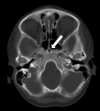
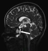
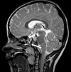
Identification of the cause of recurrent meningitis may pose a diagnostic challenge. Evaluation of a patient with recurrent meningitis calls for meticulous review of skull base structures by cross sectional imaging to exclude any underlying anatomical abnormality. Our case highlights the importance of excluding persistent craniopharyngeal duct, a rare but treatable cause of recurrent meningitis. The isolation of Streptococcus pneumoniae in recurrent meningitis may be a clue to the presence of a skull base abnormality. Craniopharyngeal canals have been classified depending on their qualitative and quantitative imaging features. Such imaging based classification is important for identification of patients with associated potential pituitary involvement and also for appropriate surgical planning. Controversy exists as to the approach to surgical treatment of craniopahryngeal duct. The persistent craniopahryngeal duct in our patient was successfully treated by an endoscopic transsphenoidal approach.
Figures


References
-
- Hughes ML, Carty AT, White FE. . Persistent hypophyseal (craniopharyngeal) canal. Br J Radiol 1999; 72: 204–6. - PubMed
-
- Madeline LA, Elster AD. . Postnatal development of the central skull base: normal variants. Radiology 1995; 196: 757–63. - PubMed
-
- Pinilla-Arias D, Hinojosa J, Esparza J, Muñoz A. . Recurrent meningitis and persistence of craniopharyngeal canal: case report. Neurocirugia (Astur) 2009; 20: 50–3. - PubMed
Publication types
LinkOut - more resources
Full Text Sources
Other Literature Sources

