

Case Reports
doi: 10.1259/bjrcr.20150399.
eCollection 2016.
Congenital hepatic haemangioma leading to multiple organ failure in a neonate
Affiliations
- PMID: 30363646
- PMCID: PMC6180865
- DOI: 10.1259/bjrcr.20150399
Item in Clipboard
Case Reports
Congenital hepatic haemangioma leading to multiple organ failure in a neonate
BJR Case Rep.
.
Abstract
We report a case of a premature male newborn who died from multiple organ failure due to a large congenital hepatic haemangioma that was diagnosed by imaging. Congenital haemangioma is a vascular tumour. The liver is the second organ involved after the skin. This tumour can be asymptomatic but can also lead to death.
Figures
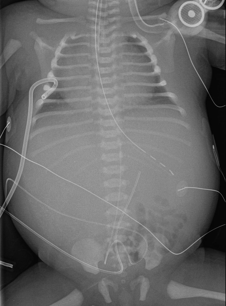
X-ray of the chest and abdomen showed cardiomegaly, acute pulmonary and soft tissues oedema, and an opacity related to hepatomegaly.
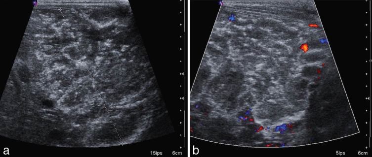
Ultrasound demonstrated a hyperechoic, heterogeneous hepatic mass (between calipers) (a) with moderate vascularization on Doppler ultrasound (b) and small calcifications.
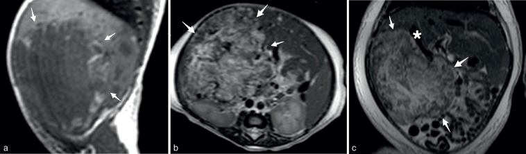
MRI showed a heterogeneous hepatic mass (arrows) in the right lobe, mostly hypointense on T
1 (a, parasagittal view) and hyperintense on T
2 weighted (b, axial view; c, frontal view) images, with enlarged hepatic veins (asterisk).
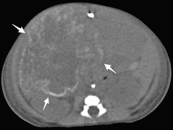
Non-enhanced CT scan showed predominantly peripheral calcifications (arrows).
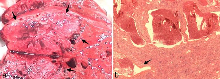
(a) Gross anatomy, large necrotic hepatic mass (arrows) of the right lobe, adjacent to normal parenchyma. (b) Histological specimen, large intratumoral necrosis with peripheral varying-sized vascular lakes (arrow). The glucose transporter-1 marker was negative.
References
-
- ISSVA Classification of Vascular Anomalies © 2014 International Society for the Study of Vascular Anomalies [Accessed April2014] Available from: issva.org/classification
-
- Franchi-Abella S, Gorincour G, Avni F, Guibaud L, Chevret L, Pariente D, et al. Hepatic haemangioma-prenatal imaging findings, complications and perinatal outcome in a case series. Pediatr Radiol 2012; 42: 298–307. - PubMed
-
- Berenguer B, Mulliken JB, Enjolras O, Boon LM, Wassef M, Josset P, et al. Rapidly involuting congenital hemangioma: clinical and histopathologic features. Pediatr Dev Pathol 2003; 6: 495–510. - PubMed
-
- Gorincour G, Kokta V, Rypens F, Garel L, Powell J, Dubois J. Imaging characteristics of two subtypes of congenital hemangiomas: rapidly involuting congenital hemangiomas and non-involuting congenital hemangiomas. Pediatr Radiol 2005; 35: 1178–85. - PubMed
-
- Dubois J, Alison M. Vascular anomalies: what a radiologist needs to know. Pediatr Radiol 2010; 40: 895–905. - PubMed
Publication types
LinkOut - more resources
Full Text Sources
