

Dorsal Root Ganglion Maintains Stemness of Bone Marrow Mesenchymal Stem Cells by Enhancing Autophagy through the AMPK/mTOR Pathway in a Coculture System
- PMID: 30363977
- PMCID: PMC6186314
- DOI: 10.1155/2018/8478953
Dorsal Root Ganglion Maintains Stemness of Bone Marrow Mesenchymal Stem Cells by Enhancing Autophagy through the AMPK/mTOR Pathway in a Coculture System
Abstract
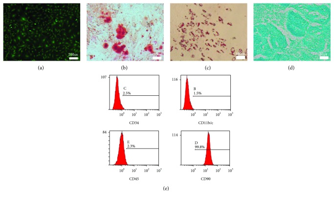
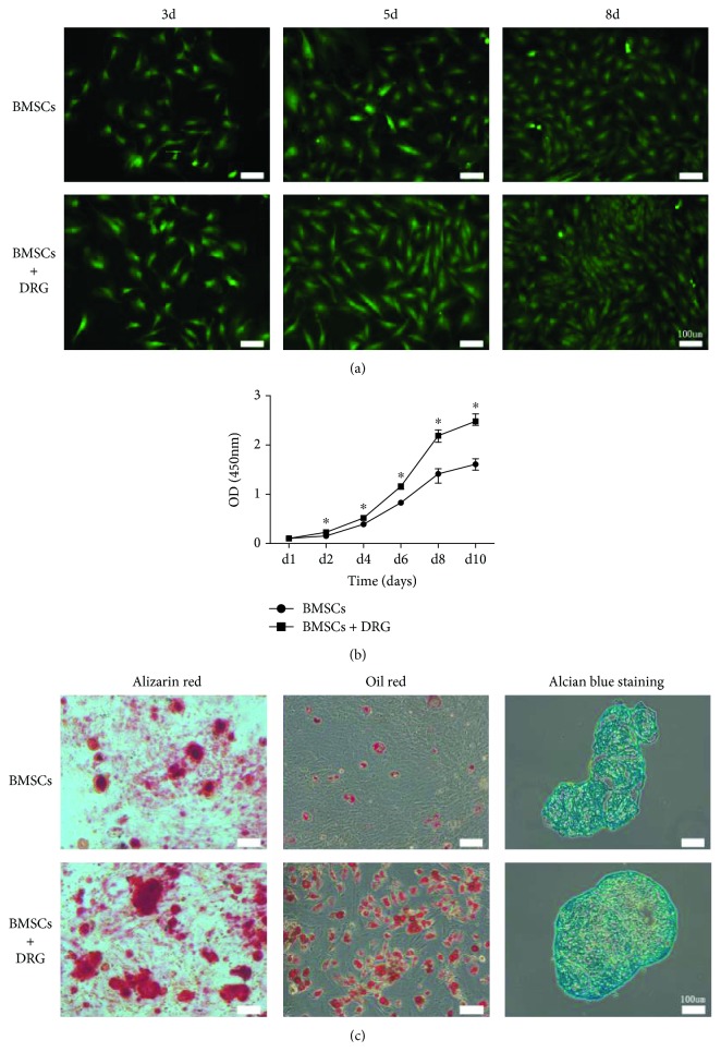
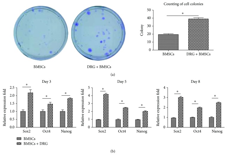
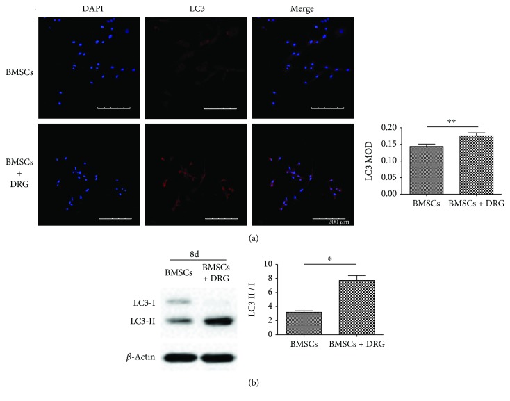
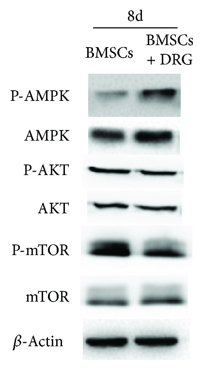
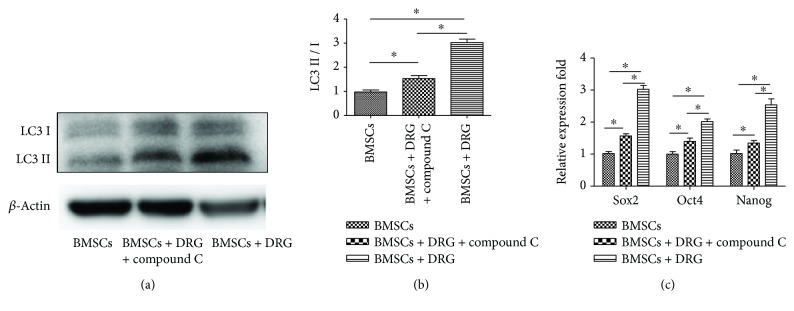
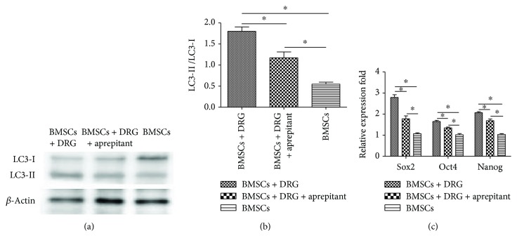
Our previous studies found that sensory nerve tracts implanted in tissue-engineered bone (TEB) could result in better osteogenesis. To explore the mechanism of the sensory nerve promoting osteogenesis in TEB in vitro, a transwell coculture experiment was designed between dorsal root ganglion (DRG) cells and bone marrow mesenchymal stem cells (BMSCs). BMSC proliferation was determined by CCK8 assay, and osteo-, chondro-, and adipogenic differentiation were assessed by alizarin red, alcian blue, and oil red staining. We found that the proliferation and multipotent differentiation of BMSCs were all enhanced in the coculture group compared to the BMSCs group. Crystal violet staining showed that the clone-forming ability of BMSCs in the coculture group was also enhanced and mRNA levels of Sox2, Nanog, and Oct4 were significantly upregulated in the coculture group. Moreover, the autophagy level of BMSCs, regulating their stemness, was promoted in the coculture group, mediated by the AMPK/mTOR pathway. In addition, AMPK inhibitor compound C could significantly downregulate the protein expression of LC3 and the mRNA level of stemness genes in the coculture group. Finally, we found that the NK1 receptor antagonist, aprepitant, could partly block this effect, which indicated that substance P played an important role in the effect. Together, we conclude that DRG could maintain the stemness of BMSCs by enhancing autophagy through the AMPK/mTOR pathway in a transwell coculture system, which may help explain the better osteogenesis after implantation of the sensory nerve into TEB.
Figures
Similar articles
-
A comparative study of HAMSCs/HBMSCs transwell and mixed coculture systems.IUBMB Life. 2019 Jul;71(7):1048-1055. doi: 10.1002/iub.2074. Epub 2019 May 21. IUBMB Life. 2019. PMID: 31112365
-
Osteogenic differentiation and angiogenesis with cocultured adipose-derived stromal cells and bone marrow stromal cells.Biomaterials. 2014 Jun;35(17):4792-804. doi: 10.1016/j.biomaterials.2014.02.048. Epub 2014 Mar 18. Biomaterials. 2014. PMID: 24655782
-
Bone marrow mesenchymal stem cells inhibit cardiac hypertrophy by enhancing FoxO1 transcription.Cell Biol Int. 2021 Jan;45(1):188-197. doi: 10.1002/cbin.11482. Epub 2020 Oct 22. Cell Biol Int. 2021. PMID: 33049085
-
[Comparative study between hypoxia and hypoxia mimetic agents on osteogenesis of bone marrow mesenchymal stem cells in mouse].Zhongguo Xiu Fu Chong Jian Wai Ke Za Zhi. 2016 Jul 8;30(7):903-908. doi: 10.7507/1002-1892.20160181. Zhongguo Xiu Fu Chong Jian Wai Ke Za Zhi. 2016. PMID: 29786329 Chinese.
-
Xeno-Free Spheroids of Human Gingiva-Derived Progenitor Cells for Bone Tissue Engineering.Front Bioeng Biotechnol. 2020 Aug 19;8:968. doi: 10.3389/fbioe.2020.00968. eCollection 2020. Front Bioeng Biotechnol. 2020. PMID: 32974308 Free PMC article.
Cited by
-
TMEM16A Activation Inhibits Autophagy in Dorsal Root Ganglion Cells, Which is Associated with the p38 MAPK/mTOR Pathway.Cell Mol Neurobiol. 2024 Dec 4;45(1):1. doi: 10.1007/s10571-024-01507-z. Cell Mol Neurobiol. 2024. PMID: 39630319 Free PMC article.
-
GPR30 Alleviates Pressure Overload-Induced Myocardial Hypertrophy in Ovariectomized Mice by Regulating Autophagy.Int J Mol Sci. 2023 Jan 4;24(2):904. doi: 10.3390/ijms24020904. Int J Mol Sci. 2023. PMID: 36674423 Free PMC article.
-
[Gene silencing of Nemo-like kinase promotes neuralized tissue engineered bone regeneration].Beijing Da Xue Xue Bao Yi Xue Ban. 2025 Apr 18;57(2):227-236. doi: 10.19723/j.issn.1671-167X.2025.02.002. Beijing Da Xue Xue Bao Yi Xue Ban. 2025. PMID: 40219550 Free PMC article. Chinese.
-
Modern Trends for Peripheral Nerve Repair and Regeneration: Beyond the Hollow Nerve Guidance Conduit.Front Bioeng Biotechnol. 2019 Nov 22;7:337. doi: 10.3389/fbioe.2019.00337. eCollection 2019. Front Bioeng Biotechnol. 2019. PMID: 31824934 Free PMC article. Review.
-
MiR-19b-3p accelerates bone loss after spinal cord injury by suppressing osteogenesis via regulating PTEN/Akt/mTOR signalling.J Cell Mol Med. 2021 Jan;25(2):990-1000. doi: 10.1111/jcmm.16159. Epub 2020 Dec 17. J Cell Mol Med. 2021. PMID: 33332749 Free PMC article.
References
-
- Pneumaticos S. G., Triantafyllopoulos G. K., Basdra E. K., Papavassiliou A. G. Segmental bone defects: from cellular and molecular pathways to the development of novel biological treatments. Journal of Cellular and Molecular Medicine. 2010;14(11):2561–2569. doi: 10.1111/j.1582-4934.2010.01062.x. - DOI - PMC - PubMed
LinkOut - more resources
Full Text Sources
Research Materials
Miscellaneous
