. 2018 Oct 1:2018:3082560.
doi: 10.1155/2018/3082560. eCollection 2018.

Treatment of Diabetic Macular Edema with Intravitreal Antivascular Endothelial Growth Factor and Prompt versus Deferred Focal Laser during Long-Term Follow-Up and Identification of Prognostic Retinal Markers

Affiliations

Treatment of Diabetic Macular Edema with Intravitreal Antivascular Endothelial Growth Factor and Prompt versus Deferred Focal Laser during Long-Term Follow-Up and Identification of Prognostic Retinal Markers

Birgit Weingessel et al. J Ophthalmol. .

Abstract

Purpose: Long-term follow-up of patients with diabetic macular edema (DME) treated with intravitreal antivascular endothelial growth factor (anti-VEGF) combined focal laser and identification of prognostic morphological characteristics.

Methods: Prospective clinical trial (50 treatment-naive eyes) with DME randomized 1 : 1 receiving intravitreal ranibizumab (0.5 mg/0.05 ml) and prompt grid laser compared with ranibizumab and deferred laser. Morphological characteristics potentially relevant for prognosis were assessed at baseline, month 6, month 9, and years 1, 2, 3, 4, and 5 of follow-up.

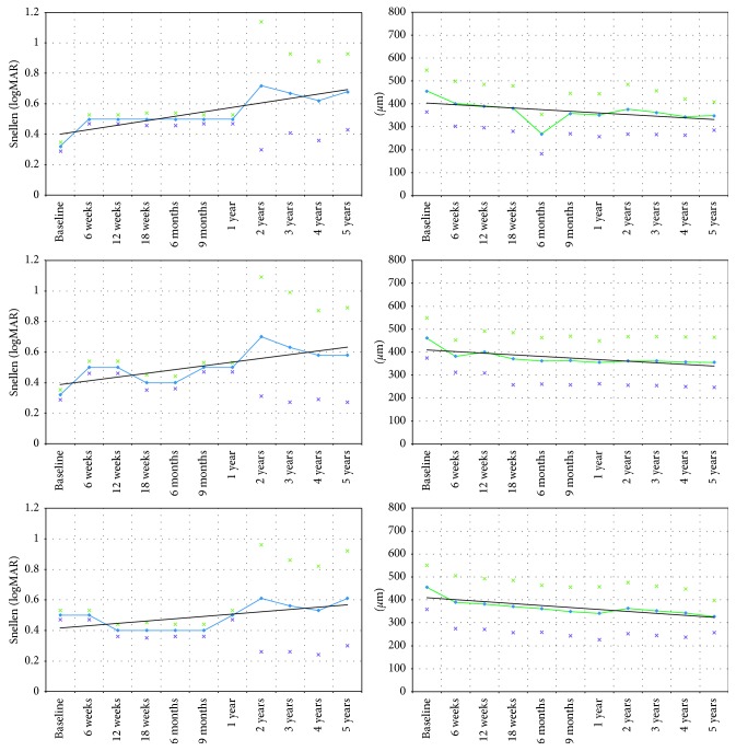
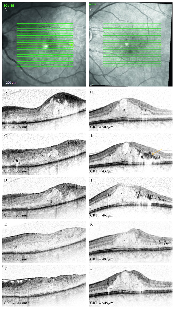
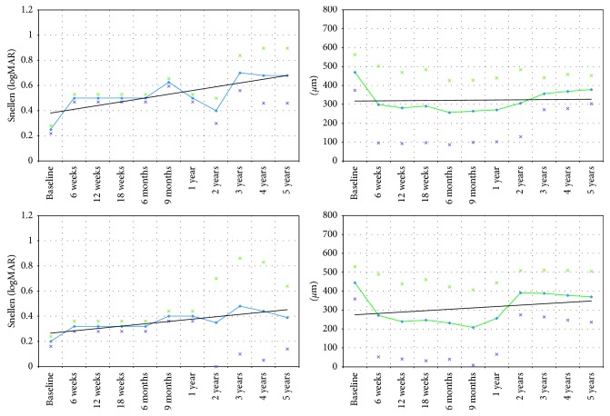
Results: Although functional results were slightly higher in the prompt group at week 12 (0.5; 20/40 Snellen (SD = 0.04, 0.3 logMAR) versus 0.4; 20/50 Snellen (SD = 0.04, logMAR: 0.4), p=0.4) and month 9 (prompt group: 0.5; 20/40 Snellen (SD = 0.03, 0.3 logMAR) versus deferred group: 0.4; 20/50 Snellen (SD = 0.04, 0.4 logMAR), p=0.4), these were statistically insignificant. There was no significant benefit regarding functionality during long-term follow-up in the prompt group compared to the deferred group. BCVA in the eyes with clusters of hyperreflective foci in the central macular region was inferior compared with the eyes without these alterations at year 5 (0.39; 20/50 Snellen, (SD = 0.25, 0.4 logMAR) versus 0.63; 20/80 Snellen (SD = 0.22, 0.2 logMAR), p < 0.01).

Conclusion: Grid laser and ranibizumab therapy are effective in DME management during the long-term follow-up. Intraretinal hyperreflective material in SD-OCT is negatively related to BCVA.


Figures

Figure 1
Figure 1
Best-corrected visual acuity (BCVA) and central retinal thickness (CRT) in eyes with diabetic macular edema (DME) treated with intravitreal antivascular endothelial growth factor (anti-VEGF) combined with prompt or deferred focal macular laser. Although functional results in eyes treated with immediate anti-VEGF therapy combined with laser at week 12 (prompt treatment group: 0.5; 20/40 Snellen (SD = 0.04, 0.3 logMAR) versus deferred treatment group: 0.4; 20/50 Snellen (SD = 0.04, logMAR: 0.4), p=0.4) and month 9 (prompt group: 0.5; 20/40 Snellen (SD = 0.03, 0.3 logMAR) versus deferred group: 0.4; 20/50 Snellen (SD = 0.04, 0.4 logMAR), p=0.4)) were slightly better; no meaningful functional benefit was observed in the prompt group during long-term follow-up. BCVA = best-corrected visual acuity. CRT = central retinal thickness. Black lines represent trend lines. Stars represent standard deviation. p values indicate change compared with baseline. (a) BCVA-entire study population (50 eyes). (b) CRT-entire study population (50 eyes). (c) BCVA-prompt/deferred (light blue line) group (25/25 eyes). (d) CRT-prompt/deferred (light green line) group (25/25 eyes).
Figure 2
Figure 2
Spectral domain optical coherence tomography (SD-OCT) images of the left eye of 2 patients treated with ranibizumab and prompt focal laser therapy (A–F) or ranibizumab in addition to deferred laser (H–L) at baseline (B, H), 6 months (C, I), 1 year (D, J), 2 years (E, K), and 5 years (F, L) following baseline. SD-OCT images indicate a more rapid and consistent decrease of central retinal thickness (CRT) values in the eye treated with prompt laser therapy compared with the eye randomized for deferred focal laser treatment. In this example, the decrease in CRT is particularly evident in the eye that received prompt laser therapy in the beginning of the treatment phase (until month 6) compared with the eye that was scheduled for the deferred group. Note that the baseline CRT was similar in both eyes of the different treatment arms. The arrow indicates clusters of hyperreflective intraretinal material.
Figure 3
Figure 3
Best-corrected visual acuity (BCVA) and central retinal thickness (CRT) in eyes with diabetic macular edema (DME) treated with intravitreal antivascular endothelial growth factor (anti-VEGF) and focal macular laser. Data represent eyes revealing clusters of intraretinal hyperreflective material as detected on spectral domain optical coherence tomography (SD-OCT) and eyes without these morphological changes. BCVA in eyes with clusters of hyperreflective foci in the central macular region detected by SD-OCT was significantly inferior compared with eyes without intraretinal hyperreflective foci at year 5 of follow-up (0.39; 20/50 Snellen (SD = 0.25, 0.4 logMAR) versus 0.63; 20/32 Snellen (SD = 0.22, 0.2 logMAR), p < 0.01). BCVA = best-corrected visual acuity. CRT = central retinal thickness. Black lines represent trend lines. Stars indicate standard deviation. p values indicate change compared with baseline. (a) BCVA in eyes with (22 eyes) and without (20 eyes, light blue line) intraretinal hypherreflective foci in SD-OCT. (b) CRT in eyes with (22 eyes) and without (28 eyes, light green line) intraretinal hypherreflective foci in SD-OCT.

Similar articles

Cited by

References

    1. Klein R., Klein B. E., Moss S. E., Davis M. D., DeMets D. L. The wisconsin epidemiologic study of diabetic retinopathy. IV: diabetic macular edema. Ophthalmology. 1984;91(12):1464–1474. doi: 10.1016/s0161-6420(84)34102-1. - DOI - PubMed
    1. Moss S. E., Klein R., Klein B. E. The 14-year incidence of visual loss in a diabetic population. Ophthalmology. 1998;105(6):998–1003. doi: 10.1016/s0161-6420(98)96025-0. - DOI - PubMed
    1. Stewart M. W. A review of ranibizumab for the treatment of diabetic retinopathy. Ophthalmology and Therapy. 2017;6(1):33–47. doi: 10.1007/s40123-017-0083-9. - DOI - PMC - PubMed
    1. Funatsu H., Yamashita H., Sakata K., et al. Vitreous levels of vascular endothelial growth factor and intracellular adhesion molecule 1 are related to diabetic macular edema. Ophthalmology. 2005;112(5):806–816. doi: 10.1016/j.ophtha.2004.11.045. - DOI - PubMed
    1. Nathan D. M., Genuth S., Lachin J., et al. Photocoagulation therapy for diabetic eye disease: Early Treatment Diabetic Retinopathy Study Research Group. JAMA. 1985;254(21):3086–3087. - PubMed

LinkOut - more resources