. 2018 Oct 10:6:134.
doi: 10.3389/fcell.2018.00134. eCollection 2018.

The 3-Phosphoinositide-Dependent Protein Kinase 1 Inhibits Rod Photoreceptor Development

Affiliations

The 3-Phosphoinositide-Dependent Protein Kinase 1 Inhibits Rod Photoreceptor Development

Tiaosi Xing et al. Front Cell Dev Biol. .

Abstract

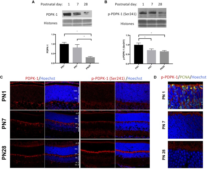
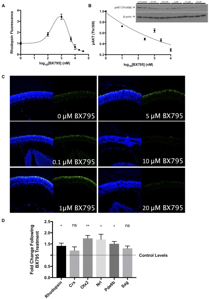
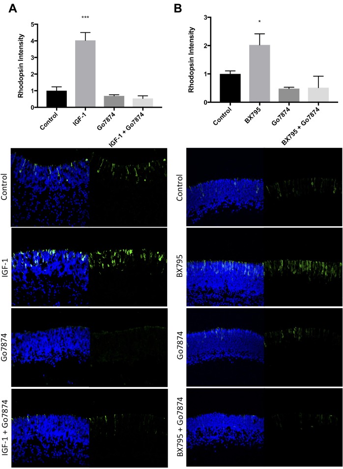
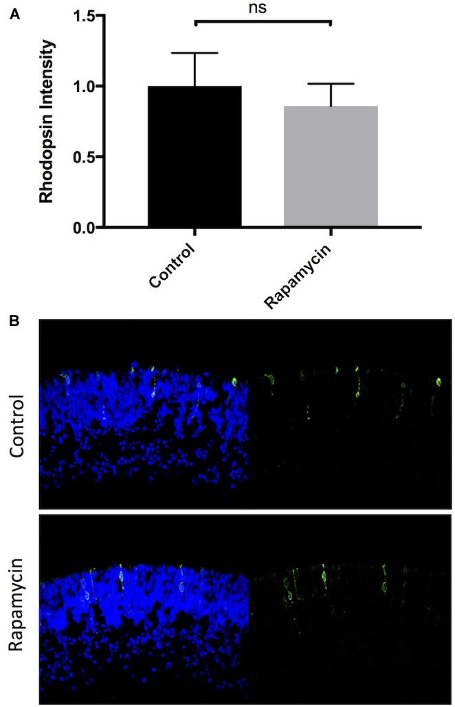
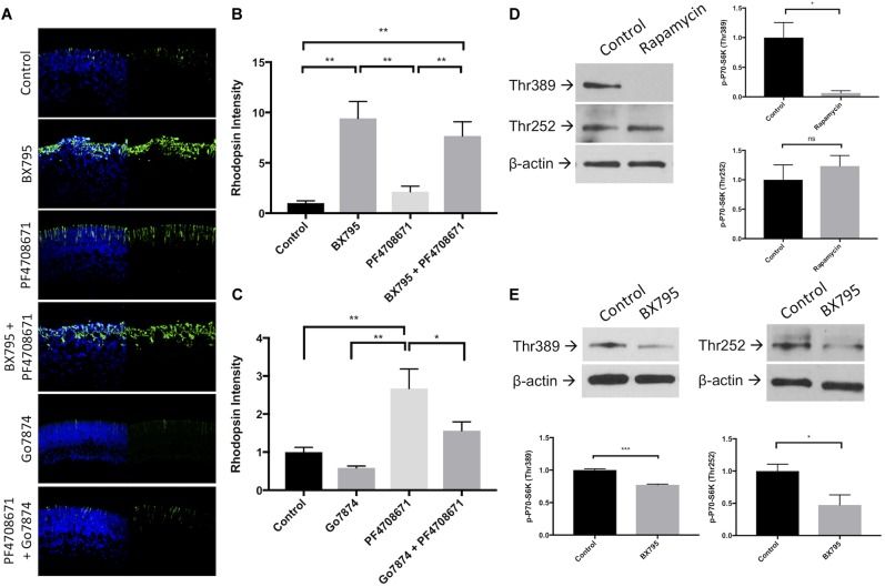
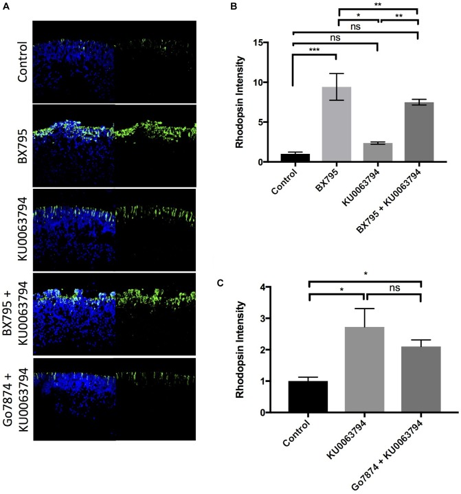
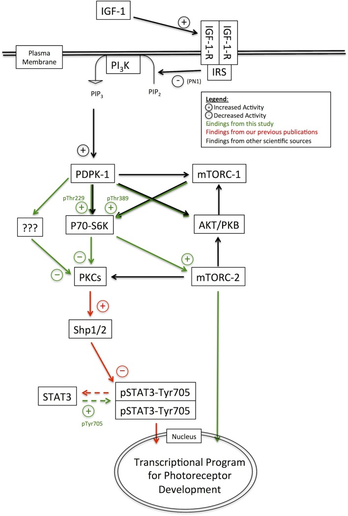
The transition of rod precursor cells to post-mitotic rod photoreceptors can be promoted by extrinsic factors such as insulin-like growth factor 1 (IGF-1), which regulates phosphatidylinositide concentration, and consequently the 3-phosphoinositide-dependent protein kinase-1 (PDPK-1). PDPK-1 is a 63 kDa cytoplasmic kinase that controls cell proliferation and differentiation. In the mouse retina, PDPK-1 and its phosphorylated derivative p-PDPK-1 (Ser241), showed peak expression during the first postnatal (PN) day with a substantial decline by PN7 and in the adult retina. Though initially widely distributed among cell types, PDPK-1 expression decreased first in the inner retina and later in the outer retina. When PDPK-1 is inhibited in neonatal retinal explants by BX795, there is a robust increase in rod photoreceptor numbers. The increase in rods depended on the activity of PKC, as BX795 had no effect when PKC is inhibited. Inhibition of PDPK-1-dependent kinases, such as P70-S6K, but not others, such as mTORC-1, stimulated rod development. The P70-S6K-dependent increase in rods appears to be correlated with phosphorylation of Thr252 and not at Thr389, a substrate of mTORC-1. This pathway is also inactive while PKC activity is inhibited. We also found that inhibition of the kinase mTORC-2, also stimulated by insulin activity, similarly increased rod formation, and this effect appears to be independent of PKC activity. This may represent a novel intracellular signaling pathway that also stimulates photoreceptor development. Consistent with previous studies, stimulation of STAT3 activity is sufficient to prevent any PDPK-1, P70-S6K, or mTORC2-dependent increase in rods. Together the data indicate that PDPK-1 and other intrinsic kinases downstream of IGF-1 are key regulators of rod photoreceptor formation.

Keywords: IGF-1; PDPK-1; PKC; STAT3; development; mTORC2; photoreceptor; retina.


Figures

FIGURE 1
FIGURE 1
PDPK-1 and p-PDPK-1 expression decline following retinal development. Western blots and corresponding histograms representing (A) PDPK-1 and (B) p-PDPK-1 Ser241 levels in wild-type PN 1, 7 and adult mouse retinas, normalized to the three histone bands on a coomassie stain of the gel. (C) Immunofluorescent labeling of PDPK-1 and p-PDPK-1 Ser241 (red) and Hoechst-33258 (blue) in 10 μm sections of the postnatal days 1, 7, and 28 wild-type mouse retina. (D) The same set of retinas, fluorescently labeled for p-PDPK-1 (red), PCNA (green), and Hoechst-33258 (blue). In this set of images, the objective is focused on the outer neuroblast or nuclear layers. ONBL, outer neuroblast layer; INBL, inner neuroblast layer; OS, outer segments; ONL, outer nuclear layer; OPL, outer plexiform layer; INL, inner nuclear layer; IPL, inner plexiform layer; GCL, ganglion cell layer. n = 3 biological replicates/measure p < 0.05, ∗∗p < 0.01, and ∗∗∗p < 0.001.
FIGURE 2
FIGURE 2
BX795 is a PDPK-1 inhibitor that enhances rod photoreceptor precursor development. In P1 retinal explant cultures, PDPK-1 was inhibited with 0, 0.1, 1, 5, 10, 20, and 40 μM BX795. (A) Quantification of rhodopsin labeling intensity as a function of log10 (BX795 concentration), relative to untreated controls. (B) Protein levels of p-AKT1Thr308 as a function of BX795 concentration. The inset contains an example western blot with duplicate lanes for each concentration of BX795 (n = 3). (C) Representative images for the curve in A, 96 h retinal explants were sectioned and labeled for rhodopsin (green) and the DNA stain Hoechst-33258 (blue). (D) cDNA levels corresponding to the expression of photoreceptor and photoreceptor precursor-related genes. n = 3 biological replicates/measure. p < 0.05 and ∗∗p < 0.01.
FIGURE 3
FIGURE 3
IGF-1 and BX795 enhance rod development in a PKC-dependent manner. Quantification (top) and representative images (bottom) of rhodopsin (green) and Hoechst-33258 (blue) labeled retinal explants following a 96-h exposure to nothing (controls) or (A) 50 nM IGF-1, 100 nM Go7874, or both; (B) 100 nM Go7874, 100 nM BX795, or both. n = 3 biological replicates/measure. p < 0.05 and ∗∗∗p < 0.001.
FIGURE 4
FIGURE 4
Inhibition of mTORC-1 does not affect rod photoreceptor development. Quantification and representative images of rhodopsin (green) and Hoechst-33258 (blue) labeled retinal explants following a 96-h exposure to nothing (controls) or 10 nM rapamycin.
FIGURE 5
FIGURE 5
Inhibition of P70-S6K with PF4708671 enhances rod photoreceptor development. (A) Representative rhodopsin (green) and Hoechst-33258 (blue) labeling in retinal explants following a 96 h exposure to nothing (control), 100 nM BX795, 200 nM PF4708671, BX795 and PF4708671, 100 nM Go7874, or Go7874 and PF4708671. (B,C) Quantification of treatment groups represented in (A). (D) Example western blots and densitometric analysis of retinal explants left untreated or treated with 10 nM Rapamycin for 30 min. Lysate from these retinas was labeled with antibodies to detect P70-S6K Thr252, P70-S6K Thr389, and β-actin. (E) The same proteins were detected in lysate from retinal explants treated with 100 nM BX795 for 30 min. n = 3 biological replicates/measure. p < 0.05, ∗∗p < 0.01, and ∗∗∗p < 0.001.
FIGURE 6
FIGURE 6
Inhibition of mTORC-1/2 with KU0063794 enhances rod photoreceptor development independently of PKC. (A) Example images and (B,C) quantification of rhodopsin labeling in retinal explants following a 96-h exposure to nothing (controls) or (B) 100 nM KU0063794, 100 nM BX795, or both; (C) 100 nM KU0063794, or both. n = 3 biological replicates/measure. p < 0.05, ∗∗p < 0.01, and ∗∗∗p < 0.001.
FIGURE 7
FIGURE 7
STAT3 activation suppresses rod photoreceptor development, which is prevented by BX795. (A) Quantification and (B) example western blot demonstrating the effects of 30 min exposures to BX795, PF4708671, and KU0063794 on STAT3 Tyr705 phosphorylation in retinal explants. (C) Quantification and (D) representative images of rhodopsin (green) and Hoechst-33258 (blue)-labeled retinas following a 96 h exposure to nothing (control), 100 nM BX795, 100 nM KU0063794, 200 nM PF4708671 (left column), or any of the above in combination with 20 ng/mL LIF (right column). p < 0.05, ∗∗p < 0.01, and ∗∗∗p < 0.001.
FIGURE 8
FIGURE 8
Pathway diagram representing the pathways and interacting factors that control rod photoreceptor development.

References

    1. Alajajian B. B., Fletcher L., Isgor E., Jimenez D. F., Digicaylioglu M. (2009). IGF-I regulated phosphorylation and translocation of PDK-1 in neurons. Neuroreport 20 579–583. 10.1097/WNR.0b013e328329a41a - DOI - PubMed
    1. Alessi D. R., Andjelkovic M., Caudwell B., Cron P., Morrice N., Cohen P., et al. (1996). Mechanism of activation of protein kinase B by insulin and IGF-1. EMBO J. 15 6541–6551. 10.1002/j.1460-2075.1996.tb01045.x - DOI - PMC - PubMed
    1. Alessi D. R., Downes C. P. (1998). The role of PI 3-kinase in insulin action. Biochim. Biophys. Acta 1436 151–164. 10.1016/S0005-2760(98)00133-7 - DOI - PubMed
    1. Alessi D. R., James S. R., Downes C. P., Holmes A. B., Gaffney P. R., Reese C. B., et al. (1997). Characterization of a 3-phosphoinositide-dependent protein kinase which phosphorylates and activates protein kinase Balpha. Curr. Biol. 7 261–269. 10.1016/S0960-9822(06)00122-9 - DOI - PubMed
    1. Andjelković M., Alessi D. R., Meier R., Fernandez A., Lamb N. J., Frech M., et al. (1997). Role of translocation in the activation and function of protein kinase B. J. Biol. Chem. 272 31515–31524. 10.1074/jbc.272.50.31515 - DOI - PubMed

LinkOut - more resources