Review
. 2018 Oct 10:8:345.
doi: 10.3389/fcimb.2018.00345. eCollection 2018.

Cellular and Molecular Immune Response to Chikungunya Virus Infection

Affiliations
Review

Cellular and Molecular Immune Response to Chikungunya Virus Infection

Ithallo S B Tanabe et al. Front Cell Infect Microbiol. .

Abstract

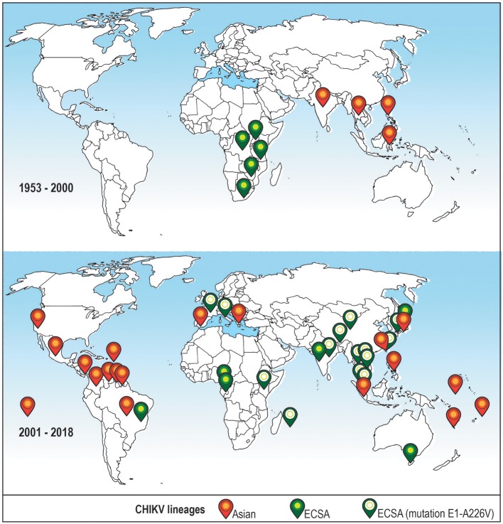
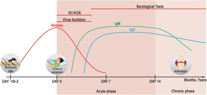
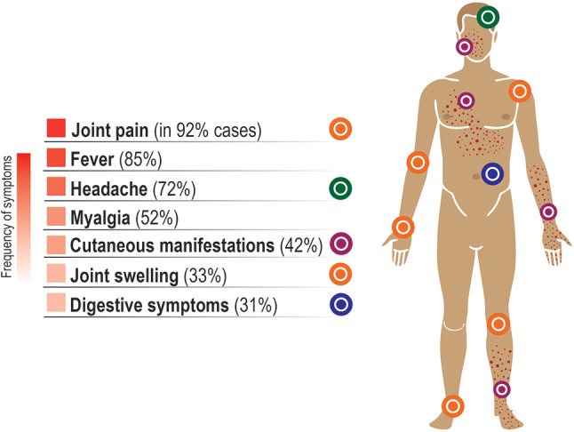
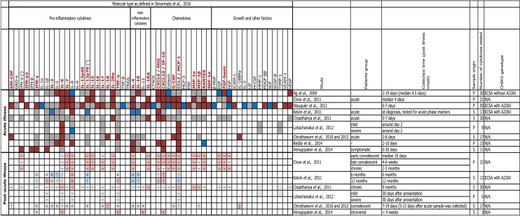
Chikungunya virus (CHIKV) is a re-emergent arthropod-borne virus (arbovirus) that causes a disease characterized primarily by fever, rash and severe persistent polyarthralgia. In the last decade, CHIKV has become a serious public health problem causing several outbreaks around the world. Despite the fact that CHIKV has been around since 1952, our knowledge about immunopathology, innate and adaptive immune response involved in this infectious disease is incomplete. In this review, we provide an updated summary of the current knowledge about immune response to CHIKV and about soluble immunological markers associated with the morbidity, prognosis and chronicity of this arbovirus disease. In addition, we discuss the progress in the research of new vaccines for preventing CHIKV infection and the use of monoclonal antibodies as a promising therapeutic strategy.

Keywords: Chikungunya virus; adaptative immunity; immune response; immunological markers; immunovirology; innate immunity; vaccines.


Figures

Figure 1
Figure 1
Distribution of CHIKV lineages that are associated with recent outbreaks around the world before and after year 2000. Top: CHIKV cases reported in the XX century (1953–2000). Bottom: CHIKV cases reported recently (2001–2018). Only cases where the virus lineage was identified are shown. Orange icon–Asian lineage; green icon–East/Central/South African (ECSA) lineage; green icon with a white circle–ECSA strain with a mutation A226V in the E1 envelope glycoprotein, this strain is sometimes referred to as Indian Ocean lineage (Wahid et al., 2017).
Figure 2
Figure 2
Applicability of different diagnostic methods in the course of CHIKV infection. In the acute phase, viremia can persist until days 5–7 pio (Silva and Dermody, 2017) and CHIKV genomic RNA can be detected by RT-PCR reliably until day 7 pio (Edwards et al., 2017). It is therefore suggested that the detection of CHIKV RNA and virus isolation from serum samples for diagnostic purposes is done before day 5 pio (Johnson et al., 2016b), because the chance of false-negative results increases with the decrease in viral load. IgM and IgG antibodies against CHIKV begin to be produced at days 2 pio (Jain et al., 2018) and 4 pio (Prince et al., 2015), respectively. Stable titers of IgM can be seen in the serum from day 6 pio till around 4 months pio (Prince et al., 2015) [and can be detected mostly until 6 months pio (Chua et al., 2017)], whereas sustained levels of IgG can be present for more than 1 year (Chua et al., 2017). The antibodies against CHIKV can be detected by immunoassays after the development of humoral immune response (in case of IgG–long into the chronic phase, both–symptomatic or asymptomatic). A more detailed overview of the methods available for diagnostics of CHIKV is given in a review by Sam et al. (2015).
Figure 3
Figure 3
Symptoms of the acute phase of CHIKV infection. Some minor variation exists in the frequency of symptoms reported in different studies. Typically, the clinical symptoms in the acute phase of the disease include high fever, pain and swelling in the joints, myalgia, and skin rash, often accompanied by headache, backache and fatigue. Here, the average percentage of symptomatic cases where a given symptom was reported is based on the data from Thiberville et al. (2013b), with the exception for the percentages for fever and headache that were taken from Huits et al. (2018). These symptoms usually remain for about 5–7 days as a self-limiting disease and are followed by a complete recovery within 2 weeks. However, severe joint pain can remain for months or even years in some individuals, often in distal joints (Roosenhoff et al., 2016) and in fluctuating manner (Hoarau et al., 2010). It has been estimated that ~16% of cases are asymptomatic (Thiberville et al., 2013b). The symptoms in CHIKV-infected children differ from those in adults and are listed in Table 1 from the study by Simarmata et al. (2016).
Figure 4
Figure 4
Profile of pro- and anti-inflammatory cytokines, chemokines and growth factors in human CHIKV infection. The heatmap shows soluble immune mediators that were measured in patients during the acute and post-acute phases (where available) of CHIKVD in ten studies. For comparisons between CHIKV-infected and healthy individuals, dark and light colors are used to highlight the differences or lack of thereof during the acute and post-acute phases, correspondingly; red indicates significant upregulation, blue–significant downregulation, gray–lack of significant differences in the levels of a given cytokine/chemokine/growth factor, and white indicates that those levels were not measured. For comparisons between acute and post-acute phases, ↓ and ↑ symbols indicate down- and upregulation in the post-acute phase, × symbol indicates the absence of significant differences, and “–” (en dash) symbol indicates that no comparison was reported. Where not specified, we assumed that: IL-12 is IL12p70 ('), and IFN-α is IFN-α1 (''). Patients group names are given as in the corresponding study. Sample origin: S–serum, P–plasma. CHIKV genotype: ECSA–East/Central/South African, N/A–no data. Names of the factors that are shown to be age-independently upregulated in the acute phase of CHIKVD by Simarmata et al. (2016) are shown in dark red. References: (Ng et al., ; Chirathaworn et al., , ; Chaaitanya et al., ; Chow et al., ; Kelvin et al., ; Wauquier et al., ; Lohachanakul et al., ; Reddy et al., ; Venugopalan et al., 2014).

References

    1. Abdelnabi R., Neyts J., Delang L. (2015). Towards antivirals against chikungunya virus. Antiviral Res. 121, 59–68. 10.1016/j.antiviral.2015.06.017 - DOI - PMC - PubMed
    1. Agarwal A., Sharma A. K., Sukumaran D., Parida M., Dash P. K. (2016). Two novel epistatic mutations (E1:K211E and E2:V264A) in structural proteins of Chikungunya virus enhance fitness in Aedes aegypti. Virology 497, 59–68. 10.1016/j.virol.2016.06.025 - DOI - PubMed
    1. Ahola T., Merits A. (2016). Functions of Chikungunya virus nonstructural proteins, in Chikungunya Virus: Advances in Biology, Pathogenesis, and Treatment, ed Okeoma C. M. (Cham: Springer International Publishing; ), 75–98.
    1. Akahata W., Yang Z. Y., Andersen H., Sun S., Holdaway H. A., Kong W. P., et al. . (2010). A virus-like particle vaccine for epidemic Chikungunya virus protects nonhuman primates against infection. Nat. Med. 16, 334–338. 10.1038/nm.2105 - DOI - PMC - PubMed
    1. Althouse B. M., Guerbois M., Cummings D. A. T., Diop O. M., Faye O., Faye A., et al. . (2018). Role of monkeys in the sylvatic cycle of chikungunya virus in Senegal. Nat. Commun. 9, 1046. 10.1038/s41467-018-03332-7 - DOI - PMC - PubMed

Publication types

MeSH terms