

Complications After Tibial Tuberosity Osteotomy: Association With Screw Size and Concomitant Distalization
- PMID: 30364433
- PMCID: PMC6196632
- DOI: 10.1177/2325967118803614
Complications After Tibial Tuberosity Osteotomy: Association With Screw Size and Concomitant Distalization
Abstract
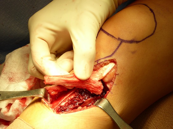
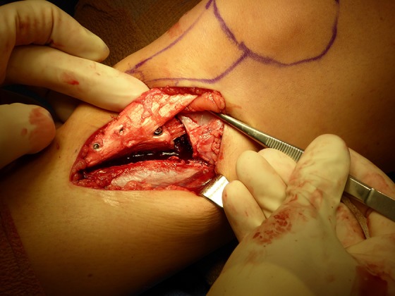
Background: Tibial tuberosity osteotomy (TTO) is a versatile procedure commonly used to treat patellar instability as well as to unload cartilage lesions. TTO with concomitant distalization (TTO-d) may be performed in patients with patella alta to stabilize the patella by helping it to engage in the trochlea earlier during flexion.
Purpose: To identify and compare perioperative complications in patients who underwent TTO and those who underwent TTO-d and to analyze risk factors associated with these complications.
Study design: Cohort study; Level of evidence, 3.
Methods: We retrospectively identified perioperative complications and associated factors from medical records for 240 patients who underwent TTO with or without distalization performed by 2 surgeons at 2 institutions between 2009 and 2015. A musculoskeletal radiologist at each institution determined osteotomy union using a published grading system. Significance was set at P < .01.
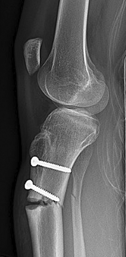
Results: Of the 240 patients, 153 (122 TTO, 31 TTO-d) had clinical and radiographic follow-up of at least 90 days or evidence of osseous union. Eighty-eight complications were identified in 71 of 153 (46%) patients: delayed union (n = 35); painful hardware (n = 32); deep vein thrombosis (n = 4); clinical nonunion, delayed range of motion, sensory deficit, and wound breakdown (n = 3 each); and broken screw, fascial hernia, hematoma, quadriceps dysfunction, and tibial fracture (n = 1 each). Thirteen of 35 delayed unions occurred in the TTO-d group (P = .005). Painful hardware was more frequent in patients who received 4.5-mm screws (31/115) than in those who received 3.5-mm screws (1/38) (P = .001). A reoperation was required in 38 of 153 patients (37 patients using 4.5-mm screws vs 1 patient using 3.5-mm screws; P < .001), primarily for screw removal (32/38).
Conclusion: Minor complications, including delayed union and painful hardware, were common, but major complications such as tibial fracture, deep vein thrombosis, and clinical nonunion were rare. Delayed union was more frequent in the TTO-d group. The 3.5-mm screws were less painful and less likely to need removal than the 4.5-mm screws.
Keywords: anteromedialization; complications; dislocation; distalization; instability; osteotomy; patella; patellofemoral; tibia; tuberosity.
Conflict of interest statement
One or more of the authors has declared the following potential conflict of interest or source of funding: D.N.M. is a paid section editor for Rheumatology. B.E.S.S. is a consultant for Arthrex, is a paid speaker/presenter for Arthrex, and has received hospitality payments from DePuy. A.J.C. has received research support from the Arthroscopy Association of North America, has been paid to provide expert testimony, and receives royalties from Elsevier. AOSSM checks author disclosures against the Open Payments Database (OPD). AOSSM has not conducted an independent investigation on the OPD and disclaims any liability or responsibility relating thereto.
Figures
References
-
- Bellemans J, Cauwenberghs F, Brys P, Victor J, Fabry G. Fracture of the proximal tibia after Fulkerson anteromedial tibial tubercle transfer: a report of four cases. Am J Sports Med. 1998;26(2):300–302. - PubMed
-
- Bhandari M, Chiavaras M, Ayeni O, et al. Assessment of radiographic fracture healing in patients with operatively treated femoral neck fractures. J Orthop Trauma. 2013;27(9):e213–e219. - PubMed
-
- Fulkerson JP. Anteromedialization of the tibial tuberosity for patellofemoral malalignment. Clin Orthop Relat Res. 1983;177:176–181. - PubMed
-
- Fulkerson JP, Becker GJ, Meaney JA, Miranda M, Folcik MA. Anteromedial tibial tubercle transfer without bone graft. Am J Sports Med. 1990;18(5):490–496. - PubMed
LinkOut - more resources
Full Text Sources
