Case Reports
. 2016 Jan 19;2(1):20150275.
doi: 10.1259/bjrcr.20150275. eCollection 2016.

Acute massive congestive ischaemic colitis related to inferior mesenteric arteriovenous malformation

Affiliations
Case Reports

Acute massive congestive ischaemic colitis related to inferior mesenteric arteriovenous malformation

Bruno Coulier et al. BJR Case Rep. .

Abstract

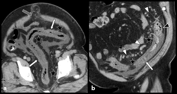
We report a very rare case of acute congestive ischaemic colitis of the left colon caused by brutal decompensation of an uncommon arteriovenous malformation (AVM) in the territory of the inferior mesenteric artery (IMA) in a 45-year-old male patient. The patient presented with severe abdominal pain in the left iliac fossa and abundant mucoid stools. The diagnosis of congestive colitis was made by optical colonoscopy but the full diagnosis of the responsible AVM in the IMA territory was made by contrast-enhanced multidetector CT scan combined with colour Doppler ultrasound. Two successive attempts at selective embolization failed to resolve the symptoms and finally, extensive surgery was necessary. The complete imaging findings of the case are presented and the characteristic features of uncommon AVMs and fistulas of the IMA territory are briefly reviewed.


Figures

Figure 1.
Figure 1.
Optical colonoscopy (a, b) of the sigmoid colon shows the extremely oedematous, cobblestone appearance of the colonic mucosa causing narrowing of the lumen, especially at the level of the descending colon (b). No ulceration or blood was found.
Figure 2.
Figure 2.
Contrast-enhanced abdominal CT images. Oblique (a) and coronal oblique (b) multiplanar reconstructions show severe but homogeneously circumferential hypodense thickening of a rather long segment of the left colon extending from the descending colon to the rectosigmoid junction (white arrows) and fat stranding of the umbilical hernia (grey arrow). There is a sharp cutoff of the thickening at the level of the left phrenicocolic ligament. Diffuse congestive fat stranding is associated with the mesosigmoid and the greater omentum (black stars). Unusually large and serpiginous varicosities are found not only in the mesosigmoid fat (white arrowheads on b) but also in the colonic wall (black arrowhead on b). Ascites are visible in the sigmoid and the right iliac fossa.
Figure 3.
Figure 3.
Contrast-enhanced abdominal CT images. Selective volume rendering reconstruction of the massive serpiginous mesosigmoid varicosities (a). They converge to a dilated vein running through the transverse mesocolon to penetrate the venous splenoportal confluence at right angle (black arrow). An accessory venous drainage also runs to the left renal vein (grey arrow). The inferior mesenteric vein is constitutionally absent in this patient (not illustrated). The inferior mesenteric artery is also visible (white arrowheads). Complementary colour Doppler ultrasound was performed (b). Ultrasound palpation of the left iliac fossa was very painful and confirmed a very thickened hypoechoic sigmoid colon (white arrows) running through a diffusely painful and uncompressible hyperechoic inflammatory mesocolonic fat (white asterisk). Colour Doppler confirmed major varicosities not only along the thickened colonic wall (full circle) but also within the colonic wall (dotted circle). The Doppler spectrum of these varicosities contains a pulsatile arterial component (white star) strongly suggesting an arteriovenous communication.
Figure 4.
Figure 4.
Contrast-enhanced abdominal CT images. Details of the maximum intensity projection view (a) and the volume rendering view (b) show the superior rectal artery (white arrows), the normal superior rectal vein (black arrows) and the presence of an indisputable arteriovenous communication (white circles). It is noteworthy that the caliber of the rectal vein abruptly increases just distal to this anomaly (grey arrow on b). Moreover, the caliber of the inferior mesenteric artery also appears considerably increased (white arrow on d) when compared with a view obtained 4 years ago (white arrow on c).
Figure 5.
Figure 5.
Selective arteriography (a–c) of the inferior mesenteric artery confirms the diagnosis of an arteriocapillary type malformation (black arrows) with a rather low flow. Careful embolization with ethylene vinyl alcohol copolymer (ONYX®, Micro Therapeutic Inc., Irvine, CA) was performed (white arrows on d and e). Nevertheless, 3 days later, important symptoms persisted. A new angio-CT scan with volume rendering views (e) confirmed the persistence of an active arteriovenous fistula (white arrowheads).
Figure 6.
Figure 6.
Gross anatomy of the resected specimen (a, b) reveals not only extremely diffuse congestion of the sigmoid (black star) but also of the entire mesosigmoid and the epiploic appendages (white star). Transverse section through the specimen (c) shows congestion of the mesocolon with varicosities and major submucosal oedema (black arrows). Photomicrograph (haematoxylin–eosin stain; magnification, ×25) confirmed major submucosal oedema (white stars). The mucosa itself was normal (white arrow); black arrow points to the muscular layer.

References

    1. Metcalf DR, Nivatvongs S, Andrews JC. Ischemic colitis: an unusual case of inferior mesenteric arteriovenous fistula causing venous hypertension. Report of a case. Dis Colon Rectum 2008; 51: 1422–4. - PubMed
    1. Washington C, Carmichael JC. Management of ischemic colitis. Clin Colon Rectal Surg 2012; 25: 228–35. - PMC - PubMed
    1. Poullos PD, Thompson AC, Holz G, Edelman LA, Jeffrey RB. Ischemic colitis due to a mesenteric arteriovenous malformation in a patient with a connective tissue disorder. J Radiol Case Rep 2014; 8: 9–21. - PMC - PubMed
    1. Justaniah AI, Molgaard C, Flacke S, Barto A, Iqbal S. Congenital inferior mesenteric arteriovenous malformation presenting with ischemic colitis: endovascular treatment. J Vasc Interv Radiol 2013; 24: 1761–3. - PubMed
    1. Gorospe EC, Leggett CL, Sun G. Inferior mesenteric arteriovenous malformation: an unusual cause of ischemic colitis. Ann Gastroenterol 2012; 25: 165. - PMC - PubMed

Publication types