. 2019 Jan;54(1):98-110.
doi: 10.3892/ijo.2018.4604. Epub 2018 Oct 24.

Betulinic acid inhibits stemness and EMT of pancreatic cancer cells via activation of AMPK signaling

Affiliations

Betulinic acid inhibits stemness and EMT of pancreatic cancer cells via activation of AMPK signaling

Liankang Sun et al. Int J Oncol. 2019 Jan.

Abstract

Cancer stem cells (CSCs), which are found in various types of human cancer, including pancreatic cancer, possess elevated metastatic potential, lead to tumor recurrence and cause chemoradiotherapy resistance. Alterations in cellular bioenergetics through the regulation of 5' adenosine monophosphate‑activated protein kinase (AMPK) signaling may be a prerequisite to stemness. Betulinic acid (BA) is a well‑known bioactive compound with antiretroviral and anti‑inflammatory potential, which has been reported to exert anticancer effects on various types of cancer, including pancreatic cancer. The present study aimed to investigate whether BA could inhibit pancreatic CSCs via regulation of AMPK signaling. The proliferation of pancreatic cancer cells was examined by MTT and colony formation assays. The migratory and invasive abilities of pancreatic cancer cells were assessed using wound‑scratch and Transwell invasion assays. In addition, the expression levels of candidate genes were measured by reverse transcription‑quantitative polymerase chain reaction and western blotting. The results revealed that BA inhibited the proliferation and tumorsphere formation of pancreatic cancer cells, suppressed epithelial‑mesenchymal transition (EMT), migration and invasion, and reduced the expression of three pluripotency factors [SRY‑box 2 (Sox2), octamer‑binding protein 4 (Oct4) and Nanog]. Furthermore, immunohistochemical analysis confirmed that there was a significant inverse association between the expression levels of phosphorylated (P)‑AMPK and Sox2 in pancreatic cancer, and it was revealed that BA may activate AMPK signaling. Notably, knockdown of AMPK reversed the suppressive effects of BA on EMT and stemness of pancreatic cancer cells. In addition, BA reversed the effects of gemcitabine on stemness and enhanced the sensitivity of pancreatic cancer cells to gemcitabine. Collectively, these results indicated that BA may effectively inhibit pluripotency factor expression (Sox2, Oct4 and Nanog), EMT and the stem‑like phenotype of pancreatic cancer cells via activating AMPK signaling. Therefore, BA may be considered an attractive therapeutic candidate and an effective inhibitor of the stem‑like phenotype in pancreatic cancer cells. Further investigation into the development of BA as an anticancer drug is warranted.

Keywords: betulinic acid; pancreatic cancer; cancer stem cells; epithelial-mesenchymal transition; 5' adenosine monophosphate-activated protein kinase; gemcitabine.


Figures
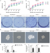
Figure 1
Figure 1
Effects of BA on the proliferation and tumorsphere formation of pancreatic cancer cells. (A and B) Mia PaCa-2 and Panc-1 cells were incubated with a series of BA concentrations (0, 12.5, 25, 50, 100 and 200 µM) for 24, 48 and 72 h, and cell viability was evaluated by MTT assay. (C and E) Effects of BA on the colony-forming ability of Mia PaCa-2 and Panc-1 cells were assessed by colony formation assay. Images are representative of three independent experiments, and the colony number was counted and plotted. Scale bar, 1 cm. (D and F) Tumorsphere formation assay of Mia PaCa-2 and Panc-1 cells treated with or without 50 µM BA. The number of tumorspheres was counted and plotted. Magnification, ×200. *P<0.05, **P<0.01. BA, betulinic acid.
Figure 2
Figure 2
Effects of BA on the migration and invasion of pancreatic cancer cells. (A) Wound-scratch assays were conducted on Mia PaCa-2 and Panc-1 cells pretreated with or without 50 µM BA. Images were visualized at 0 and 48 h. Magnification, ×100. (B-D) Effects of BA on the invasive ability of Mia PaCa-2 and Panc-1 cells were evaluated by Matrigel invasion assay. Images are representative of three independent experiments, and the invasive cells were counted and plotted. Magnification, ×200. **P<0.01. (E and F) Mia PaCa-2 and Panc-1 cells were pretreated with BA (0, 25 and 50 µM) for 48 h, and western blot analysis was performed to assess the expression of epithelial-mesenchymal transition markers (E-cadherin and vimentin). BA, betulinic acid.
Figure 3
Figure 3
BA inhibits the stemness of pancreatic cancer cells and activates AMPK signaling. (A and B) Mia PaCa-2 and Panc-1 cells were pretreated with BA for 24 h, total RNA was extracted and reverse transcription-quantitative polymerase chain reaction was conducted to detect the expression levels of Sox2, Oct4 and Nanog. *P<0.05 and **P<0.01. (C) Mia PaCa-2 and Panc-1 cells were pretreated with or without 50 µM BA for 48 h, and western blot analysis was performed to assess the protein expression levels of master pluripotency regulators (Sox2, Oct4 and Nanog), total AMPK and P-AMPK. (D and E) Mia PaCa-2 and Panc-1 cells were treated with 50 µM BA at various time-points (0, 5, 15, 30, 60 and 120 min), and total protein was extracted to detect the expression levels of P-AMPK by western blotting. (F and G) Mia PaCa-2 and Panc-1 cells were pretreated with BA for 24 h, and immunofluorescence analysis was conducted to assess the expression and nuclear localization of Sox2 in Mia PaCa-2 and Panc-1 cells. Magnification, ×400. AMPK, 5′ adenosine monophosphate-activated protein kinase; BA, betulinic acid; Oct4, octamer-binding protein 4; P, phosphorylated; Sox2, SRY-box 2.
Figure 4
Figure 4
AMPK activation by AICAR exerts similar effects to betulinic acid on EMT and stemness of pancreatic cancer cells. (A) Representative images of immunohistochemical staining of P-AMPK and SOX2 in normal pancreatic tissues and pancreatic cancer tissues. Magnification, ×100 for the upper images and ×400 for the lower images. (B) An inverse association between P-AMPK and SOX2 expression was detected in pancreatic cancer tissues. P<0.05 by two-tailed χ2 test. (C and E) Effects of AMPK activation by AICAR (2 mM) on the invasive ability of Mia PaCa-2 and Panc-1 cells were evaluated by Matrigel invasion assay. Images are representative of three independent experiments, and the invasive cells were counted and plotted. Magnification, ×200. **P<0.01. (D and F) Tumorsphere formation assay of Mia PaCa-2 and Panc-1 cells treated with or without 2 mM AICAR. The number of tumorspheres was counted and plotted. Magnification, ×200. **P<0.01. (G and H) Mia PaCa-2 and Panc-1 cells were pretreated with or without 2 mM AICAR for 48 h, and western blot analysis was performed to assess the expression levels of master pluripotency regulators (Sox2, Oct4, and Nanog) and EMT markers (E-cadherin and vimentin). AICAR, 5-aminoimidazole-4-carboxamide 1-β-D-ribofuranoside; AMPK, 5′ adenosine monophosphate-activated protein kinase; EMT, epithelial-mesenchymal transition; Oct4, octamer-binding protein 4; P, phosphorylated; PDAC, pancreatic ductal adenocarcinoma; Sox2, SRY-box 2.
Figure 5
Figure 5
Knockdown of AMPK rescues BA-induced suppression of EMT and stemness in pancreatic cancer cells. (A) Western blotting confirmed the successful silencing of AMPK in Mia PaCa-2 and Panc-1 cells. β-actin was used as an internal loading control. (B and C) Western blot analysis suggested that silencing AMPK by siRNA reversed BA-induced inhibition of the expression of master pluripotency regulators (Sox2, Oct4 and Nanog) and EMT markers (E-cadherin and vimentin) in Mia PaCa-2 and Panc-1 cells. β-actin was used as an internal loading control. (D-F) Matrigel invasion assay revealed that silencing AMPK by siRNA reversed BA-induced suppression of the invasion of Mia PaCa-2 and Panc-1 cells. Images are representative of three independent experiments, and the invasive cells were counted and plotted. Magnification, ×200. **P<0.01. (G-I) Tumorsphere formation assay exhibited that knocking down AMPK by siRNA reversed BA-induced inhibition of tumorsphere formation in Mia PaCa-2 and Panc-1 cells. The number of tumorspheres was counted and plotted. Magnification, ×200. *P<0.05, **P<0.01. AMPK, 5′ adenosine monophosphate-activated protein kinase; BA, betulinic acid; EMT, epithelial-mesenchymal transition; Oct4, octamer-binding protein 4; si/siRNA, small interfering RNA; Sox2, SRY-box 2.
Figure 6
Figure 6
BA reverses gemcitabine-induced stemness and enhances the sensitivity of pancreatic cancer cells to gemcitabine. (A) Mia PaCa-2 and Panc-1 cells were pretreated with various concentrations of gemcitabine (0, 1 and 5 µM) for 24 h, total RNA was extracted and RT-qPCR was conducted to detect the expression levels of Sox2, Oct4 and Nanog. *P<0.05 and **P<0.01, compared with the control group. (B) Mia PaCa-2 and Panc-1 cells were pretreated with various concentrations of gemcitabine (0, 1 and 5 µM) for 24 h, and western blot analysis was performed to assess the expression levels of master pluripotency regulators (Sox2, Oct4 and Nanog). (C) Effects of BA (50 µM) on gemcitabine (5 µM) treatment-induced stemness were measured by RT-qPCR analysis. *P<0.05 and **P<0.01. (D) Effects of BA (50 µM) on gemcitabine (5 µM) treatment-induced stemness were measured by western blot analysis. (E) Effects of BA (50 µM) combined with gemcitabine (5 µM) on the colony-forming ability of Panc-1 cells. (F and G) Effects of BA (50 µM) combined with gemcitabine (5 µM) on the apoptosis of Panc-1 cells. **P<0.01, compared with the control group. 7-AAD, 7-aminoactinomycin D; BA, betulinic acid; FITC, fluorescein isothiocyanate; Oct4, octamer-binding protein 4; RT-qPCR, reverse transcription-quantitative polymerase chain reaction; Sox2, SRY-box 2.

Similar articles

Cited by

References

    1. Siegel RL, Miller KD, Jemal A. Cancer statistics, 2016. CA Cancer J Clin. 2016;66:7–30. doi: 10.3322/caac.21332. - DOI - PubMed
    1. Gillen S, Schuster T, Meyer Zum, Büschenfelde C, Friess H, Kleeff J. Preoperative/neoadjuvant therapy in pancreatic cancer: A systematic review and meta-analysis of response and resection percentages. PLoS Med. 2010;7:e1000267. doi: 10.1371/journal.pmed.1000267. - DOI - PMC - PubMed
    1. Siegel R, Ma J, Zou Z, Jemal A. Cancer statistics, 2014. CA Cancer J Clin. 2014;64:9–29. doi: 10.3322/caac.21208. - DOI - PubMed
    1. Kamisawa T, Isawa T, Koike M, Tsuruta K, Okamoto A. Hematogenous metastases of pancreatic ductal carcinoma. Pancreas. 1995;11:345–349. doi: 10.1097/00006676-199511000-00005. - DOI - PubMed
    1. Gattinoni L, Klebanoff CA, Restifo NP. Paths to stemness: Building the ultimate antitumour T cell. Nat Rev Cancer. 2012;12:671–684. doi: 10.1038/nrc3322. - DOI - PMC - PubMed

MeSH terms