

Alterations of autophagy in knee cartilage by treatment with treadmill exercise in a rat osteoarthritis model
- PMID: 30365059
- PMCID: PMC6257837
- DOI: 10.3892/ijmm.2018.3948
Alterations of autophagy in knee cartilage by treatment with treadmill exercise in a rat osteoarthritis model
Abstract
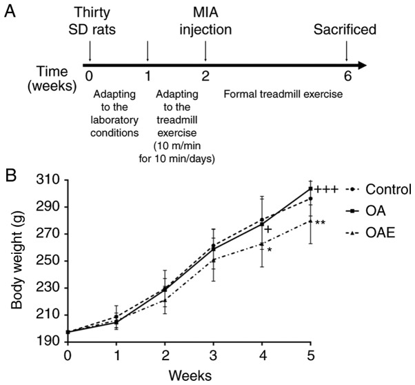
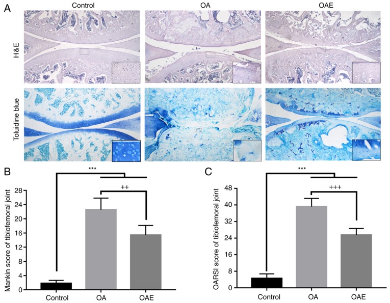
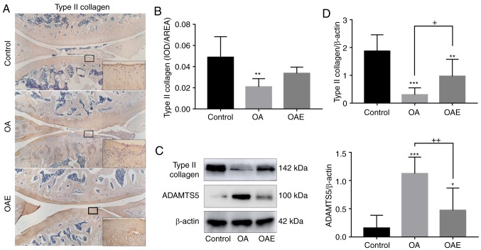
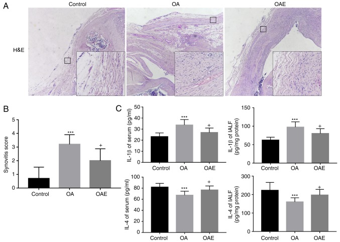
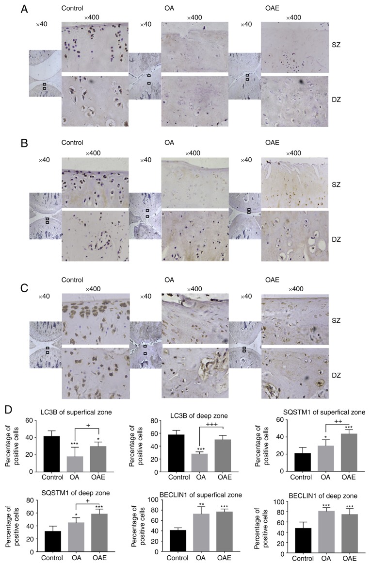
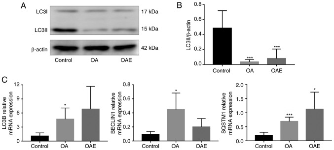
The aim of the present study was to investigate potential alterations in the articular cartilage in a rat model of monosodium iodoacetate (MIA)‑induced osteoarthritis (OA) with or without treatment with moderate treadmill exercise. A total of 30 male Sprague‑Dawley rats were randomly divided into three groups (n=10), including the control, OA and OA with treadmill exercise (OAE) groups. Rats were evaluated upon completing the treadmill exercise program (speed, 18 m/min; 30 min/day; 5 days/week for 4 weeks). Interleukin (IL)‑1β and IL‑4 levels in the serum and intra‑articular lavage fluid (IALF) were measured by ELISA. Alterations in articular cartilage and synovium were also evaluated by histology, immunohistochemistry, western blotting and reverse transcription‑quantitative polymerase chain reaction. The results revealed that IL‑1β in the serum and IALF decreased in the OAE group, whereas IL‑4 increased, and histological evaluation indicated that the OAE group had a clear treatment response. However, the expression of type II collagen in the articular cartilage increased in the OAE group as compared with the OA group, whereas ADAMTS5 expression decreased. In contrast to light chain 3B (LC3B), the protein expression levels of BECLIN1 and sequestosome 1 (SQSTM1) were increased in the OA group. In addition, a significant increase was observed between OA and OAE groups in LC3B and SQSTM1 protein levels, whereas no change was observed in BECLIN1 levels between the OA and OAE groups in the superficial and deep zones. The results of western blotting demonstrated that LC3II was notably decreased in the OA group and partially increased in the OAE group. The mRNA expression levels of LC3B and SQSTM1 increased in the OA and OAE groups, with a significant difference observed between the two groups, while a concomitant decrease was detected in BECLIN1 levels. In conclusion, 30 min of treadmill exercise had an evident protective effect in the articular cartilage of rats with MIA‑induced OA and may promote autophagy in the articular cartilage.
Figures
Similar articles
-
The effects of different frequency treadmill exercise on lipoxin A4 and articular cartilage degeneration in an experimental model of monosodium iodoacetate-induced osteoarthritis in rats.PLoS One. 2017 Jun 8;12(6):e0179162. doi: 10.1371/journal.pone.0179162. eCollection 2017. PLoS One. 2017. PMID: 28594958 Free PMC article.
-
The therapeutic effects of lipoxin A4 during treadmill exercise on monosodium iodoacetate-induced osteoarthritis in rats.Mol Immunol. 2018 Nov;103:35-45. doi: 10.1016/j.molimm.2018.08.027. Epub 2018 Sep 6. Mol Immunol. 2018. PMID: 30196232
-
Maresin-1 suppresses IL-1β-induced MMP-13 secretion by activating the PI3K/AKT pathway and inhibiting the NF-κB pathway in synovioblasts of an osteoarthritis rat model with treadmill exercise.Connect Tissue Res. 2021 Sep;62(5):508-518. doi: 10.1080/03008207.2020.1780218. Epub 2020 Jun 16. Connect Tissue Res. 2021. PMID: 32546009
-
Osteoarthritis biomarker responses and cartilage adaptation to exercise: A review of animal and human models.Scand J Med Sci Sports. 2019 Aug;29(8):1072-1082. doi: 10.1111/sms.13435. Epub 2019 May 23. Scand J Med Sci Sports. 2019. PMID: 31033061 Review.
-
Mass Spectrometry Imaging as a Potential Tool to Investigate Human Osteoarthritis at the Tissue Level.Int J Mol Sci. 2020 Sep 3;21(17):6414. doi: 10.3390/ijms21176414. Int J Mol Sci. 2020. PMID: 32899238 Free PMC article. Review.
Cited by
-
Lymphocyte and dendritic cell response to a period of intensified training in young healthy humans and rodents: A systematic review and meta-analysis.Front Physiol. 2022 Nov 11;13:998925. doi: 10.3389/fphys.2022.998925. eCollection 2022. Front Physiol. 2022. PMID: 36439269 Free PMC article.
-
Effect of moderate exercise on osteoarthritis.EFORT Open Rev. 2023 Mar 14;8(3):148-161. doi: 10.1530/EOR-22-0119. EFORT Open Rev. 2023. PMID: 36916731 Free PMC article. Review.
-
Treadmill Exercise after Controlled Abnormal Joint Movement Inhibits Cartilage Degeneration and Synovitis.Life (Basel). 2021 Apr 1;11(4):303. doi: 10.3390/life11040303. Life (Basel). 2021. PMID: 33915911 Free PMC article.
-
Unbiased comparison and modularization identify time-related transcriptomic reprogramming in exercised rat cartilage: Integrated data mining and experimental validation.Front Physiol. 2022 Sep 15;13:974266. doi: 10.3389/fphys.2022.974266. eCollection 2022. Front Physiol. 2022. PMID: 36187764 Free PMC article.
-
The Therapeutic Effects of Treadmill Exercise on Osteoarthritis in Rats by Inhibiting the HDAC3/NF-KappaB Pathway in vivo and in vitro.Front Physiol. 2019 Aug 20;10:1060. doi: 10.3389/fphys.2019.01060. eCollection 2019. Front Physiol. 2019. PMID: 31481898 Free PMC article.
References
-
- Barbour KE, Hootman JM, Helmick CG, Murphy LB, Theis KA, Schwartz TA, Kalsbeek WD, Renner JB, Jordan JM. Meeting physical activity guidelines and the risk of incident knee osteoarthritis: A population-based prospective cohort study. Arthritis Care Res (Hoboken) 2014;66:139–146. doi: 10.1002/acr.22120. - DOI - PMC - PubMed
MeSH terms
Substances
LinkOut - more resources
Full Text Sources
Medical
