. 2018 Oct 26;14(10):e1007737.
doi: 10.1371/journal.pgen.1007737. eCollection 2018 Oct.

Filamentation of asparagine synthetase in Saccharomyces cerevisiae

Affiliations

Filamentation of asparagine synthetase in Saccharomyces cerevisiae

Shanshan Zhang et al. PLoS Genet. .

Abstract

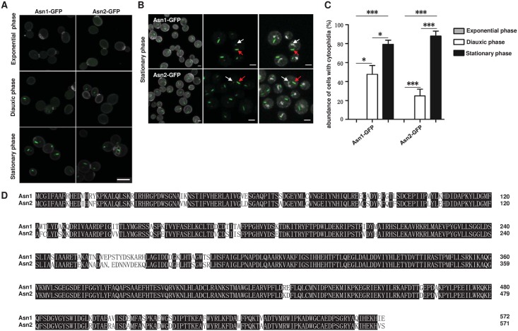
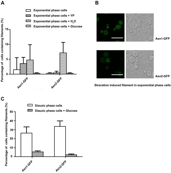
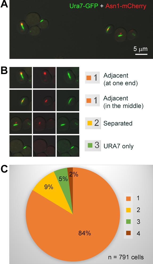
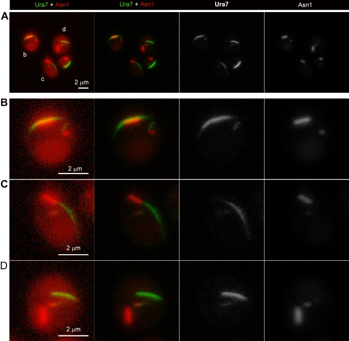
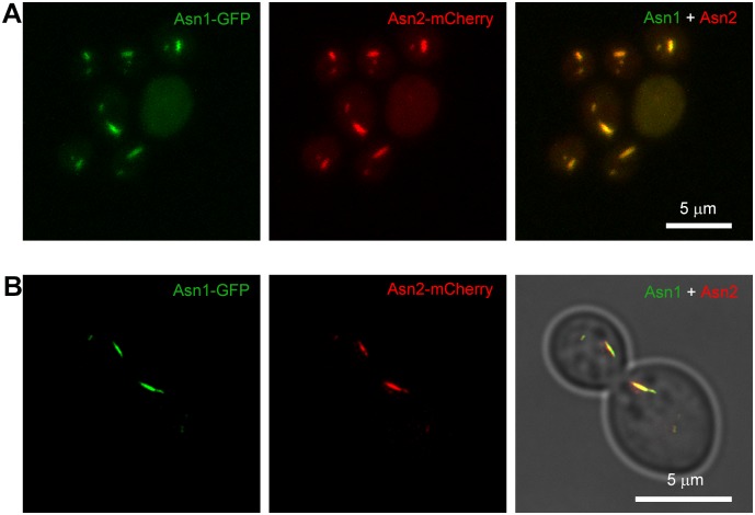
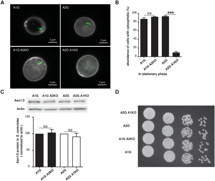
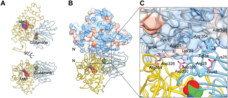
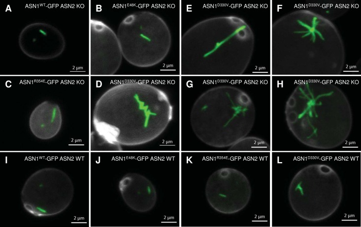
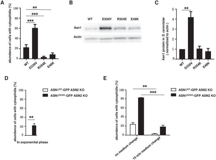
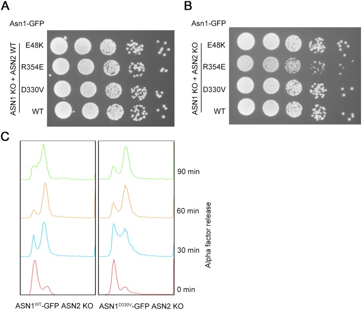
Asparagine synthetase (ASNS) and CTP synthase (CTPS) are two metabolic enzymes crucial for glutamine homeostasis. A genome-wide screening in Saccharomyces cerevisiae reveal that both ASNS and CTPS form filamentous structures termed cytoophidia. Although CTPS cytoophidia were well documented in recent years, the filamentation of ASNS is less studied. Using the budding yeast as a model system, here we confirm that two ASNS proteins, Asn1 and Asn2, are capable of forming cytoophidia in diauxic and stationary phases. We find that glucose deprivation induces ASNS filament formation. Although ASNS and CTPS form distinct cytoophidia with different lengths, both structures locate adjacently to each other in most cells. Moreover, we demonstrate that the Asn1 cytoophidia colocalize with the Asn2 cytoophidia, while Asn2 filament assembly is largely dependent on Asn1. In addition, we are able to alter Asn1 filamentation by mutagenizing key sites on the dimer interface. Finally, we show that ASN1D330V promotes filamentation. The ASN1D330V mutation impedes cell growth in an ASN2 knockout background, while growing normally in an ASN2 wild-type background. Together, this study reveals a connection between ASNS and CTPS cytoophidia and the differential filament-forming capability between two ASNS paralogs.


Conflict of interest statement

The authors have declared that no competing interests exist.

Figures

Fig 1
Fig 1. Asn1 and Asn2 form filaments in S. cerevisiae.
(A) Confocal images of yeast cells showing the filamentation capacity of Asn1 and Asn2 in exponential, diauxic and stationary phase. Scale bar, 5 μm. Asn1 and Asn2 are marked by GFP. Cell wall is stained by Calcofluor White stain. (B) In stationary phase, both Asn1 and Asn2 form cytoophidia in the cytoplasm and nucleus. Scale bar, 2 μm. The nuclei are labelled with DAPI. White arrows indicate nuclear cytoophidia and red arrows indicate cytoplasmic cytoophidia. (C) Quantification of the cells containing visible cytoophidia in the exponential, diauxic and stationary phase. *P<0.05 and ***P<0.0001. Error bars show SEM. (D) The amino acid sequence alignment of Asn1 and Asn2.
Fig 2
Fig 2. Filament formation regulated by carbon nutrient starvation.
(A, B) deprivation of glucose in media induces Asn1 and Asn2 cytoophidia in the exponential phase. (C) The addition of glucose at the diauxic shift causes Asn1 and Asn2 cytoophidia to disassemble. Error bars show 95% confidence interval. Scale bars, 10 μm. YP stands for yeast extract peptone medium without dextrose.
Fig 3
Fig 3. Association of ASNS cytoophidia and CTPS cytoophidia.
(A) Spatial relationship of ASNS cytoophidia and CTPS cytoophidia in stationary phase. Ura7 is shown in green and Asn1 is shown in red. Scale bar, 5 μm. (B) Representative images showing four kinds of spatial relationship between ASNS cytoophidia and CTPS cytoophidia. (C) Quantification of the two cytoophidia spatial relationship.
Fig 4
Fig 4. Magnified images of ASNS cytoophidia and CTPS cytoophidia in cytoplasm and nucleus.
(A) ASNS cytoophidia and CTPS cytoophidia in nucleus have similar association pattern as in cytoplasm. Scale bar, 2 μm. (B-D) High magnification of yeast cells show that the spatial relationship of ASNS cytoophidia and CTPS cytoophidia in nucleus. Scale bar, 2 μm.
Fig 5
Fig 5. Asn1 cytoophidium colocalizes with Asn2 cytoophidium.
Confocal images (A) and STED images (B) of yeast cells displayingthe colocalization of Asn1 cytoophidium and Asn2 cytoophidium. Scale bar, 5 μm. ASN1 is tagged with GFP and ASN2 is tagged with mCherry. Double tagged yeast cells are collcected in stationary phase.
Fig 6
Fig 6. Asn2 cytoophidium is dependent on Asn1.
(A) Confocal images of cytoophidium in A1G, A2G, A1G A2KO, A2G A1KO cells. Scale bar, 2 μm. (B) Quantification of cytoophidium abundance in A1G, A2G, A1G A2KO, A2G A1KO cells showing that Asn1 is crucial for Asn2 cytoophidium formation. ***P<0.0001. n.s. = not significant. Error bars show SEM. (C) Western blot analysis of ASNS protein level and quantification of the relative ASNS abundance in A1G, A2G, A1G A2KO, A2G A1KO cells. n.s. = not significant. Error bars show SEM. (D) Spot assay of A1G, A2G, A1G A2KO and A2G A1KO cells. Eight microliters of four 10-fold serial dilution of each yeast culture was spotted.
Fig 7
Fig 7. Homology model of Asn1 and its dimer.
(A) Homology model of Asn1 monomer using E.coil ASNB as reference. N-terminal domain containing the glutamine binding site and C-terminal domain containing the AMP binding site are shown in cyan and yellow, respectively. Glutamine is shown in green and AMP in magenta. (B) Homology model of Asn1 dimer. N-N’ presents Asn1 dimer interface. N’ is shown as hydrophobicity surface view. Blue represents the hydrophilic surface area and orange represents the hydrophobic surface area. (C) Asn1 dimer interface. Key residues in protein are labelled and mutation sites may have a strong effect on dimerization of Asn1 are highlighted with pink (D330, R354, E48).
Fig 8
Fig 8. Cytoophidia in Asn1 mutants.
(A-D) Morphology of Asn1WT, Asn1E48K, Asn1R354E and Asn1D330V cytoophidium in ASN2 knockout background. (E-H) Varied morphology of Asn1D330V cytoophidium in ASN1D330V-GFP ASN2 KO cells. (I-L) Morphology of Asn1WT, Asn1E48K, Asn1R354E and Asn1D330V cytoophidium in ASN2 wild type background. Scale bar, 2 μm.
Fig 9
Fig 9. Asn1 cytoophidium abundance and relative protein levels are altered in D330V mutant.
(A) The proportion of cells with Asn1 cytoophidia in different dimer interface mutants. **P<0.001. ***P<0.0001. Error bars show SEM. B) Western blot analysis of Asn1 relative protein levels. (C) Fold change of Asn1 protein level in all mutants. **P<0.001. Error bars show SEM. (D) Quantification of cytoophidium abundance of ASN1WT-GFP ASN2 KO cells and ASN1D330V-GFP ASN2 KO cells in exponential phase. **P<0.001. Error bars show SEM. (E) Effect of 15 min medium change on ASN1WT-GFP ASN2 KO cells and ASN1D330V-GFP ASN2 KO cells. **P<0.001. ***P<0.0001. Error bars show SEM.
Fig 10
Fig 10. ASN1D330V-GFP ASN2 KO cells show lower growth rate and slower cell cycle progression.
(A) Spot assay of mutants before ASN2 knockout (ASN1WT-GFP ASN2 WT, ASN1D330V-GFP ASN2 WT, ASN1R354E-GFP ASN2 WT and ASN1E48K-GFP ASN2 WT). (B) Spot assay of mutants after ASN2 knockout (ASN1WT-GFP ASN2 KO, ASN1D330V-GFP ASN2 KO, ASN1R354E-GFP ASN2 KO and ASN1E48K-GFP ASN2 KO). (C) FACS analysis of ASN1WT-GFP ASN2 KO cells and ASN1D330V-GFP ASN2 KO cells.

References

    1. Ingerson-Mahar M, Briegel A, Werner JN, Jensen GJ, Gitai Z. The metabolic enzyme CTP synthase forms cytoskeletal filaments. Nat Cell Biol. 2010;12(8):739–46. Epub 2010/07/20. 10.1038/ncb2087 . - DOI - PMC - PubMed
    1. Liu J-L. Intracellular compartmentation of CTP synthase in Drosophila. Journal of Genetics and Genomics. 2010;37(5):281–96. 10.1016/S1673-8527(09)60046-1 - DOI - PubMed
    1. Noree C, Sato BK, Broyer RM, Wilhelm JE. Identification of novel filament-forming proteins in Saccharomyces cerevisiae and Drosophila melanogaster. J Cell Biol. 2010;190(4):541–51. Epub 2010/08/18. 10.1083/jcb.201003001 . - DOI - PMC - PubMed
    1. Carcamo WC, Satoh M, Kasahara H, Terada N, Hamazaki T, Chan JY, et al. Induction of cytoplasmic rods and rings structures by inhibition of the CTP and GTP synthetic pathway in mammalian cells. PLoS ONE. 2011;6(12):e29690 Epub 2012/01/06. 10.1371/journal.pone.0029690 . - DOI - PMC - PubMed
    1. Chen K, Zhang J, Tastan OY, Deussen ZA, Siswick MY, Liu JL. Glutamine analogs promote cytoophidium assembly in human and Drosophila cells. J Genet Genomics. 2011;38(9):391–402. Epub 2011/09/21. 10.1016/j.jgg.2011.08.004 . - DOI - PubMed

Publication types

MeSH terms