. 2018 Oct 26;14(10):e1007406.
doi: 10.1371/journal.ppat.1007406. eCollection 2018 Oct.

The Citrobacter rodentium type III secretion system effector EspO affects mucosal damage repair and antimicrobial responses

Affiliations

The Citrobacter rodentium type III secretion system effector EspO affects mucosal damage repair and antimicrobial responses

Cedric N Berger et al. PLoS Pathog. .

Abstract

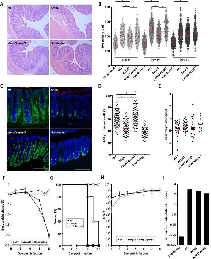
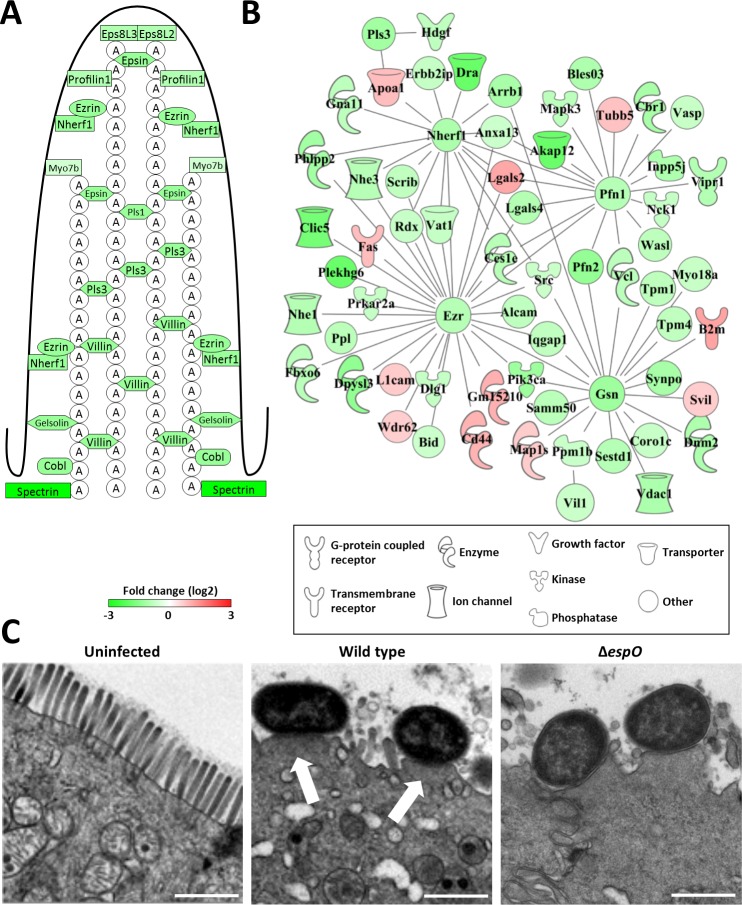
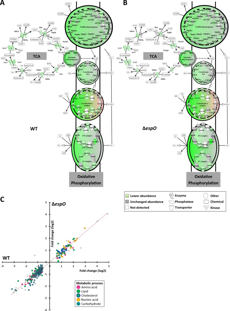
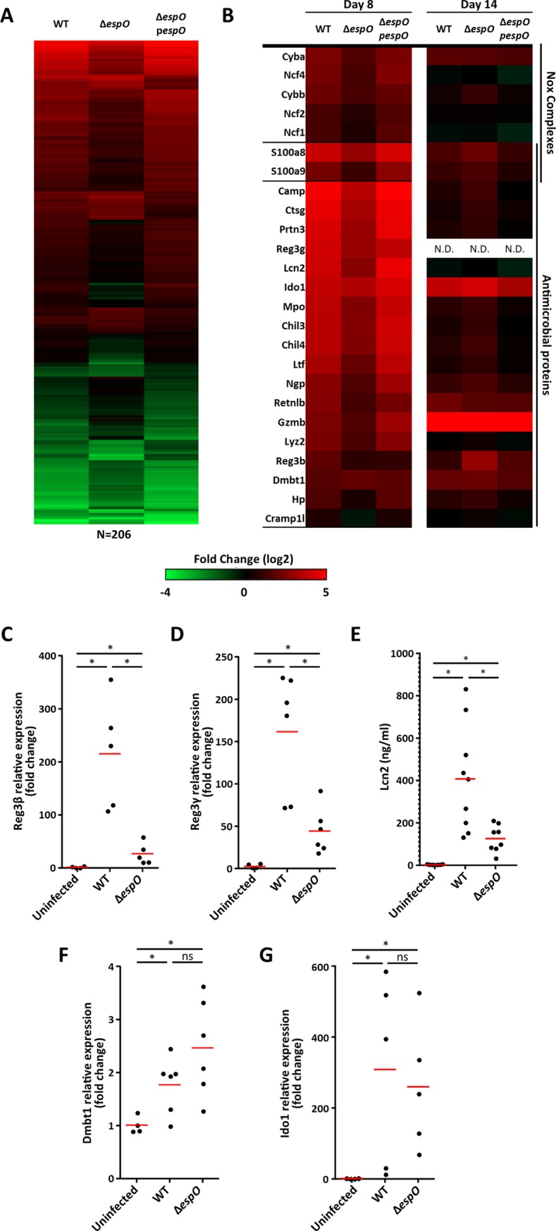
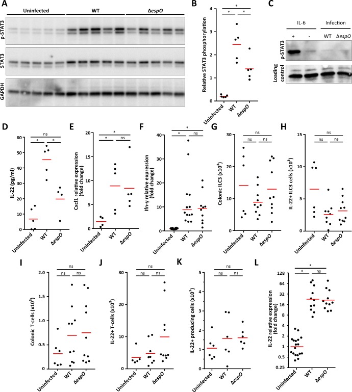
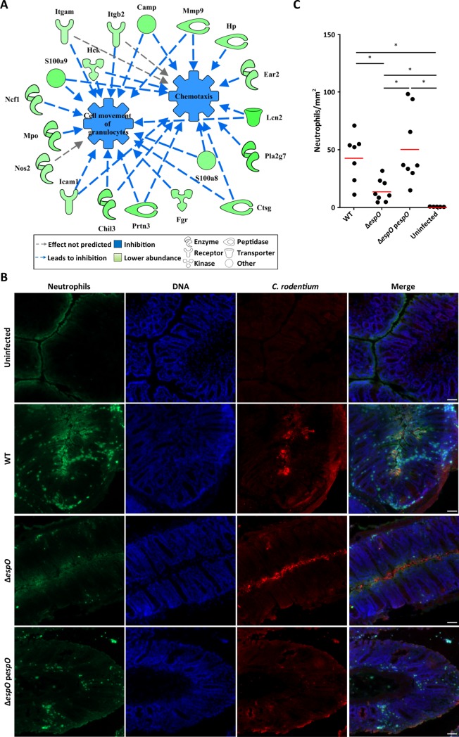
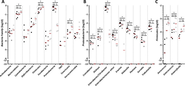
Infection with Citrobacter rodentium triggers robust tissue damage repair responses, manifested by secretion of IL-22, in the absence of which mice succumbed to the infection. Of the main hallmarks of C. rodentium infection are colonic crypt hyperplasia (CCH) and dysbiosis. In order to colonize the host and compete with the gut microbiota, C. rodentium employs a type III secretion system (T3SS) that injects effectors into colonic intestinal epithelial cells (IECs). Once injected, the effectors subvert processes involved in innate immune responses, cellular metabolism and oxygenation of the mucosa. Importantly, the identity of the effector/s triggering the tissue repair response is/are unknown. Here we report that the effector EspO ,an orthologue of OspE found in Shigella spp, affects proliferation of IECs 8 and 14 days post C. rodentium infection as well as secretion of IL-22 from colonic explants. While we observed no differences in the recruitment of group 3 innate lymphoid cells (ILC3s) and T cells, which are the main sources of IL-22 at the early and late stages of C. rodentium infection respectively, infection with ΔespO was characterized by diminished recruitment of sub-mucosal neutrophils, which coincided with lower abundance of Mmp9 and chemokines (e.g. S100a8/9) in IECs. Moreover, mice infected with ΔespO triggered significantly lesser nutritional immunity (e.g. calprotectin, Lcn2) and expression of antimicrobial peptides (Reg3β, Reg3γ) compared to mice infected with WT C. rodentium. This overlapped with a decrease in STAT3 phosphorylation in IECs. Importantly, while the reduced CCH and abundance of antimicrobial proteins during ΔespO infection did not affect C. rodentium colonization or the composition of commensal Proteobacteria, they had a subtle consequence on Firmicutes subpopulations. EspO is the first bacterial virulence factor that affects neutrophil recruitment and secretion of IL-22, as well as expression of antimicrobial and nutritional immunity proteins in IECs.


Conflict of interest statement

The authors have declared that no competing interests exist.

Figures

Fig 1
Fig 1. EspO induces CCH and cell proliferation.
(A) Representative H&E section of colonic tissue 8 DPI with WT, ΔespO or ΔespO-pespO (n = 17); uninfected mice were used as a control (scale bar 100 μm). (B) Measurements of crypt length reveal a significantly reduced level of colonic hyperplasia 8 DPI in mice infected with ΔespO compared to WT and ΔespO-pespO (*: Kruskal-Wallis test with p-value < 0.05, bars represent mean). (C) Representative immunostaining of Ki-67 (green), C. rodentium (red) and E-cadherin (blue) in colonic section from mice (n = 5) 8 DPI with ΔespO, WT or ΔespO-pespO; uninfected mice were used as a control (scale bar 100 μm). (D) Measurements of Ki-67 staining versus crypt length reveal a significantly decreased level of cell proliferation in mice infected with ΔespO compared to WT and the complemented strain (*: Kruskal-Wallis test with p-value < 0.05, bars represent mean). (E) Similar body weight was recorded for C57BL/6 mice infected with WT, ΔespO or ΔespO-pespO; uninfected mice were used as a control (n > 12). (F) Similar body weight loss was recorded in Rag2-/- il2rg-/- mice infected with WT or ΔespO; uninfected mice were used as a control (n = 5). (G) Rag2-/- il2rg-/- mice survival over time. Mice infected with ΔespO showed a small increase of survival compare to mice infected with WT C. rodentium (n = 5). (H) Fecal C. rodentium CFUs overtime. WT C. rodentium, ΔespO and ΔespO-pespO similarly colonized C57BL/6 mice up to 8 DPI. (I) Normalized abundance of IECs-associated C. rodentium proteins following infection with WT, ΔespO and the complemented strains.
Fig 2
Fig 2. The A/E lesion signature of C. rodentium infection.
(A) Schematic representation of a microvillus with structural protein abundance 8 DPI. (B) Interaction network of brush border related proteins 8 DPI. (C) TEM micrographs of uninfected and infected IECs, showing A/E lesions on 8 DPI (arrows, scale bar 1 μm).
Fig 3
Fig 3. Metabolic changes are independent of CCH.
(A) and (B) Schematic representation of the mitochondrial TCA cycle and respiratory chain showing a similar protein abundance in IECs infected with WT and ΔespO. (C) Scatter plot of the abundance (log2 fold change) of the metabolic enzyme in IEC infected with WT (x axis) and ΔespO (y axis).
Fig 4
Fig 4. EspO alters expression of AMPs.
(A) Heatmap of the 206 EspO-specific changing proteins. Abundance changes in IECs proteomes are comparing infected to uninfected mice. (B) Heatmap of differentially expressed Nox and AMPs 8 and 14 DPI. (C) and (D) Fold change in reg3β and reg3γ mRNA expression levels. (E) Fecal Lcn-2 measured by ELISA. (F) and (G) Fold change in dmbt1 and ido1 mRNA expression levels (*: Kruskal-Wallis with p-value < 0.05, each dot represents an individual mouse and bars geometric mean).
Fig 5
Fig 5. EspO triggers IL-22 release from colonic explants and STAT3 phosphorylation in IECs.
(A) Western blot showing STAT3 phosphorylation, STAT3 and GAPDH levels in IECs from uninfected, WT- and ΔespO-infected mice (5 mice/group). (B) pSTAT3 quantification (densitometry) from individual mice. (*: Kruskal-Wallis with p-value < 0.05, each dot represents and individual mouse and bars mean). (C) Western blot showing STAT3 phosphorylation levels in uninfected, WT- and ΔespO-infected HeLa cells. IL-6 was used as a positive control. No STAT3 phosphorylation was observed during infection. (D) IL-22 secretion measured by ELISA from colonic explants from uninfected, WT- and ΔespO-infected mice (*: Kruskal-Wallis with p-value < 0.05, each dot represents and individual mouse and bars mean). (E) Fold change in cxcl-1 mRNA expression level in IECs from uninfected, WT- and ΔespO-infected mice (*: Kruskal-Wallis with p-value < 0.05, each dot represents an individual mouse and bars geometric mean). (F) Fold change in ifn-γ mRNA expression level in tissue from uninfected, WT- and ΔespO-infected mice (*: Kruskal-Wallis with p-value < 0.05, each dot represents an individual mouse and bars geometric mean). (G) and (I) Bar graphs indicate the absolute numbers of colonic ILC3 (CD45.2+ CD3—CD5—CD127+ RORγt+ KLRG1—cells) and T cells (CD45.2+ CD3+ CD5+ cells). (H) and (J) Quantification of IL-22 producing ILC3 and T-cells isolated from infected colons after cytokines and PMA/Ionomycin restimulation. (K) Quantification of other IL-22 producing cells isolated from infected colons after cytokines and PMA/Ionomycin restimulation. (L) Fold change in il-22 mRNA expression level in IECs from uninfected, WT- and ΔespO-infected mice (*: Kruskal-Wallis with p-value < 0.05, each dot represents an individual mouse and bars geometric mean).
Fig 6
Fig 6. EspO promotes neutrophile transmigration.
(A) Network analysis illustrating the impact of ΔespO-regulated IECs’ proteins on chemotaxis, cell movement of granulocytes. (B) Representative immunostaining of neutrophils (Ly6G, green), C. rodentium (red) and DNA (blue) on colonic section from mice infected with WT, ΔespO and ΔespO-pespO and uninfected mice at day 8 DPI (n = 8) (scale bar 100 μm). (C) Quantification reveals a significantly decreased number of neutrophils recruited to the site of infection in mice infected with ΔespO compared to WT and the complemented strain (*: Kruskal-Wallis test with p-value < 0.05, bars represent mean).
Fig 7
Fig 7. Expression of EspO affects the composition of Firmicutes subpopulation.
(A) Changes in abundance of tissue-associated Phylum between uninfected, WT- and ΔespO-infected mice. (B) Changes in abundance of tissue-associated Proteobacteria between uninfected, WT- and ΔespO-infected mice. (C) Changes in abundance of tissue-associated Firmicutes between uninfected, WT- and ΔespO-infected mice. Each dot represents an individual mouse and bars mean (*: Kruskal-Wallis & Mann-Whitney test with FDR corrected p-value < 0.05).

Similar articles

Cited by

References

    1. Collins JW, Keeney KM, Crepin VF, Rathinam VA, Fitzgerald KA, et al. (2014) Citrobacter rodentium: infection, inflammation and the microbiota. Nat Rev Microbiol 12: 612–623. 10.1038/nrmicro3315 - DOI - PubMed
    1. Kamada N, Sakamoto K, Seo SU, Zeng MY, Kim YG, et al. (2015) Humoral Immunity in the Gut Selectively Targets Phenotypically Virulent Attaching-and-Effacing Bacteria for Intraluminal Elimination. Cell Host Microbe 17: 617–627. 10.1016/j.chom.2015.04.001 - DOI - PMC - PubMed
    1. Barthold SW (1980) The microbiology of transmissible murine colonic hyperplasia. Lab Anim Sci 30: 167–173. - PubMed
    1. Johnson E, Barthold SW (1979) The ultrastructure of transmissible murine colonic hyperplasia. Am J Pathol 97: 291–313. - PMC - PubMed
    1. Roy BC, Subramaniam D, Ahmed I, Jala VR, Hester CM, et al. (2015) Role of bacterial infection in the epigenetic regulation of Wnt antagonist WIF1 by PRC2 protein EZH2. Oncogene 34: 4519–4530. 10.1038/onc.2014.386 - DOI - PMC - PubMed

Publication types

MeSH terms

Substances

LinkOut - more resources