. 2018 Oct 26;14(10):e1007657.
doi: 10.1371/journal.pgen.1007657. eCollection 2018 Oct.

CXXC1 is not essential for normal DNA double-strand break formation and meiotic recombination in mouse

Affiliations

CXXC1 is not essential for normal DNA double-strand break formation and meiotic recombination in mouse

Hui Tian et al. PLoS Genet. .

Abstract

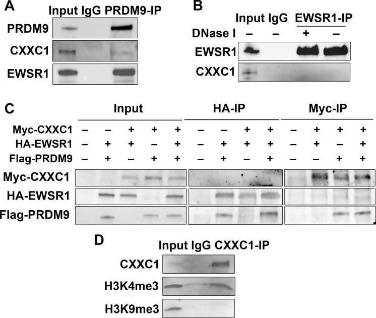
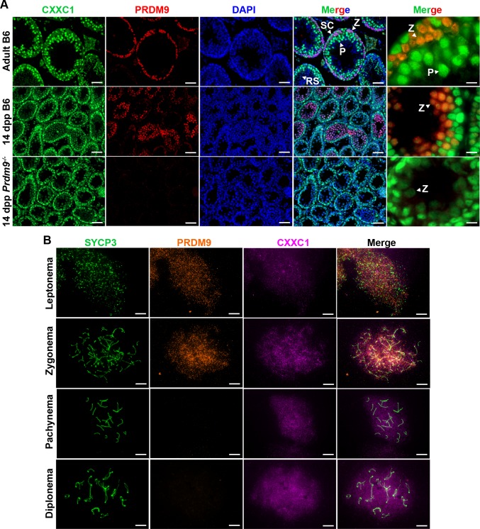
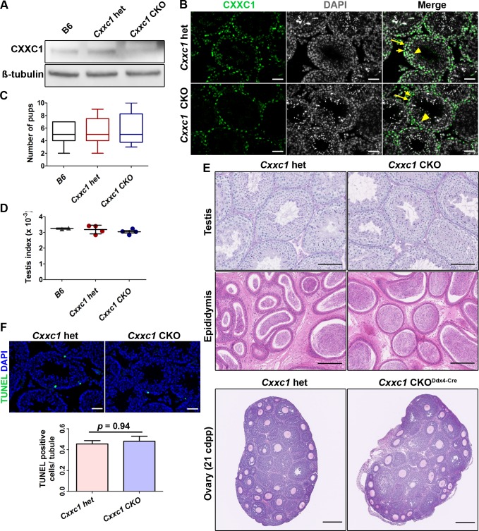
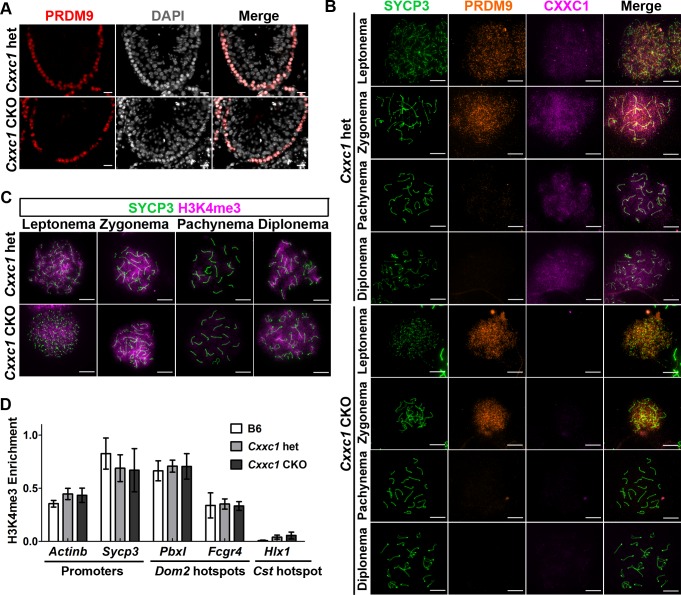
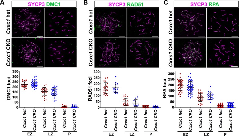
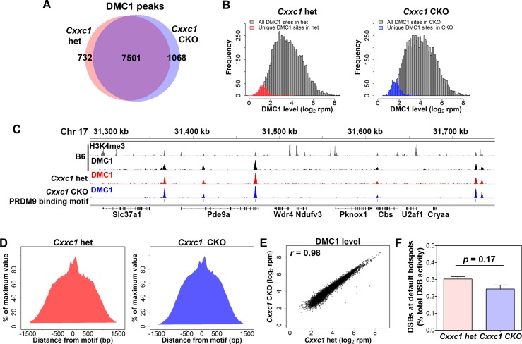
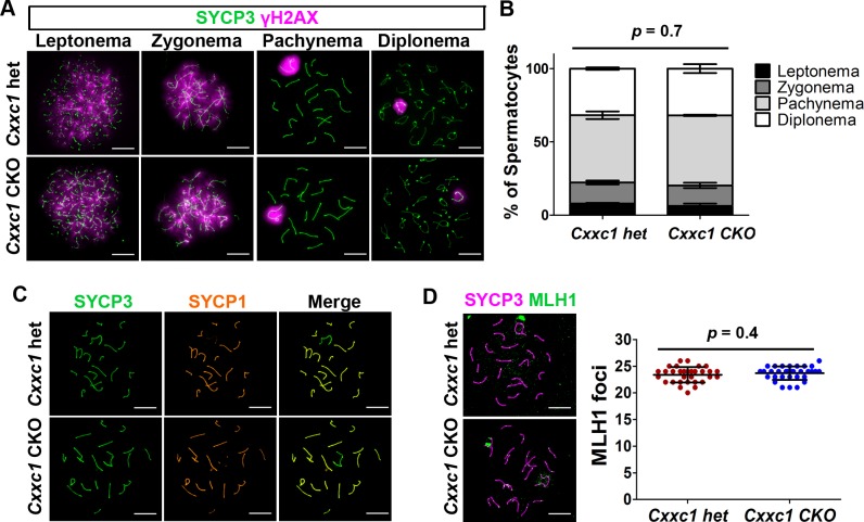
In most mammals, including mice and humans, meiotic recombination is determined by the meiosis specific histone methytransferase PRDM9, which binds to specific DNA sequences and trimethylates histone 3 at lysine-4 and lysine-36 at the adjacent nucleosomes. These actions ensure successful DNA double strand break formation and repair that occur on the proteinaceous structure forming the chromosome axis. The process of hotspot association with the axis after their activation by PRDM9 is poorly understood. Previously, we and others have identified CXXC1, an ortholog of S. cerevisiae Spp1 in mammals, as a PRDM9 interactor. In yeast, Spp1 is a histone methyl reader that links H3K4me3 sites with the recombination machinery, promoting DSB formation. Here, we investigated whether CXXC1 has a similar function in mouse meiosis. We created two Cxxc1 conditional knockout mouse models to deplete CXXC1 generally in germ cells, and before the onset of meiosis. Surprisingly, male knockout mice were fertile, and the loss of CXXC1 in spermatocytes had no effect on PRDM9 hotspot trimethylation, double strand break formation or repair. Our results demonstrate that CXXC1 is not an essential link between PRDM9-activated recombination hotspot sites and DSB machinery and that the hotspot recognition pathway in mouse is independent of CXXC1.


Conflict of interest statement

The authors have declared that no competing interests exist.

Figures

Fig 1
Fig 1. CXXC1 interacts with PRDM9 in spermatocytes.
(A) PRDM9 interacts with CXXC1 and EWSR1 in spermatocytes. Co-IP with PRDM9 from 14 dpp B6 testicular extract. Staining for each protein is indicated on left. In each blot, lane 1- input; lane 2 –co-IP with non-immune IgG; lane 3 –co-IP with antibody against PRDM9. (B) EWSR1 does not interact with CXXC1 in spermatocytes. Co-IP with EWSR1 from 14 dpp B6 testicular extract. Staining for EWSR1 and CXXC1. In each blot, lane 1- input; lane 2 –co-IP with non-immune IgG; lane 3 –co-IP with anti-EWSR1 after DNase I treatment; co-IP with anti-EWSR1 without DNase I treatment. (C) CXXC1 interacts with PRDM9 but not with EWSR1 in cell culture. Myc tagged CXXC1, HA tagged EWSR1 and Flag tagged PRDM9 were transfected into HEK293 cells. Input controls (left panel), co-IP with HA antibody (middle panel), and IP with Myc antibody (right panel). In each row, staining for the indicated proteins is shown. (D) CXXC1 binds to H3K4me3 but not to H3K9me3. Co-IP with CXXC1 from 14 dpp B6 testicular extract. Top row, staining for CXXC1; middle row, staining for H3K4me3; bottom row, staining for H3K9me3.
Fig 2
Fig 2. CXXC1 is expressed in the spermatocytes in the presence or absence of PRDM9.
(A) Immunofluorescence staining for CXXC1 and PRDM9 in adult B6, 14-dpp B6 and 14-dpp Prdm9-/- seminiferous tubule cross sections. Green, CXXC1; red, PRDM9; blue, DAPI. SC, Sertoli cell; Z, zygonema; P, pachynema; RS, round spermatid. Scale bars, first 4 columns: 50 μm, last column: 10 μm. (B) Immunofluorescence staining for CXXC1 and PRDM9 on chromosome spreads from adult B6. Green, SYCP3; orange, PRDM9; magenta, CXXC1. Scale bars, 10 μm.
Fig 3
Fig 3. Knocking out CXXC1 does not affect male fertility or testis histology.
(A) CXXC1 is depleted in Stra8-cre CKO testes. Western blot of CXXC1 from adult B6, Cxxc1 het and CKO testicular extract. ß-tubulin was used as internal loading control. (B) CXXC1 is present in Sertoli cells but not in spermatocytes of Stra8-cre CKO mice. Immunostaining of CXXC1 on Cxxc1 het and CKO seminiferous tubule cross sections. Green, CXXC1; grey, DAPI. Long arrows, Sertoli cells; short arrows, spermatogonia; arrowhead, spermatocytes. Scale bar, 50 μm. (C) No change in fertility tests in CKO mice compared to B6 and Cxxc1 heterozygous controls. The number of viable pups for each genotype is shown. (D) Testis index (testis weight/body weight) is not changed in CKO mice compared to B6 and Cxxc1 heterozygous controls. (E) Normal histology of testis, epididymis and ovary is observed in both Cxxc1 control and CKO mice. Top panels, PAS staining of seminiferous tubule sections; scale bar, 100 μm. Middle panels, H&E staining of epididymis sections; scale bar, 200 μm. Bottom panels, H&E staining of 21 dpp ovary sections, Scale bar, 250 μm. Left panels, het control; right panels, Cxxc1 CKO with Stra8-Cre in male mice, and Ddx4-Cre in females. (F) No increased apoptosis is observed in testes of CKO mice. TUNEL staining in Cxxc1 het and CKO. Top panels, scale bar, 50 μm. Bottom panels, the apoptotic cell number is quantified as TUNEL positive cell number per seminiferous tubule. Data represent as mean ± SD, p = 0.94 by Student t-test.
Fig 4
Fig 4. PRDM9 expression and catalytic function are not impaired in Cxxc1 CKO.
(A) Immunostaining of PRDM9 shows unchanged pattern in adult CKO seminiferous tubules compared to the heterozygous control. Red, PRDM9; grey, DAPI. Scale bar, 20 μm. (B) PRDM9 and SYCP3 expression patterns are not changed in CXXC1 CKO chromosome spreads compared to the heterozygous control. Co-immunostaining of CXXC1 and PRDM9 on chromosome spreads from adult Cxxc1 het and CKO mice. Green, SYCP3; orange, PRDM9; magenta, CXXC1. First 4 rows, het control; last 4 rows, Cxxc1 CKO. Scale bar, 10 μm. (C) Meiosis progression occurs normally in Cxxc1 CKO testes. Immunostaining of H3K4me3 in adult Cxxc1 het and CKO chromosome spreads. Green, SYCP3; magenta, H3K4me3. Scale bar, 10 μm. (D) Testis-specific gene expression is not changed in Cxxc1 CKO testes. H3K4me3 ChIP-qPCR with chromatin isolated form Cxxc1 het and CKO mice. Promoter regions form Actinb and Sycp3, Dom2 hotspots PbxI and Fcgr4 were amplified. Cst hotspot HlxI was used as a negative control. Bars present mean ± SD of three biological replicates.
Fig 5
Fig 5. DSB number is not affected in Cxxc1 CKO.
The DSB number was determined by three markers reflecting different stages of their processing. (A) DMC1 staining on Cxxc1 control and CKO chromosome spread. Bottom panels, distribution plot of DMC1 foci in early zygotene (n = 22 in het, n = 34 in CKO), late zygotene (n = 29 in het, n = 36 in CKO) and pachytene (n = 36 in het, n = 26 in CKO) spermatocytes. (B) RAD51 staining on Cxxc1 control and CKO chromosome spread. Bottom panels, distribution plot of RAD51 foci in early zygotene (n = 22 in het, n = 13 in CKO), late zygotene (n = 24 in het, n = 11 in CKO) and pachytene (n = 52 in het, n = 36 in CKO) spermatocytes. (C) RPA staining on Cxxc1 control and CKO chromosome spread. Bottom panels, distribution plot of RPA foci in early zygotene (n = 35 in het, n = 43 in CKO), late zygotene (n = 33 in het, n = 28 in CKO) and pachytene (n = 89 in het, n = 92 in CKO) spermatocytes. For A-C, two individuals per genotype were measured. Bars represent mean ± SD. Scale bar, 10 μm.
Fig 6
Fig 6. DSB occur at PRDM9 dependent sites in Cxxc1 CKO.
ChIP-seq data determining the number and positioning of DSB sites in CKO and heterozygous controls. (A) Venn diagram of DMC1 peak number in Cxxc1 het and CKO. 7,501 peaks are shared in Cxxc1 het and CKO. (B) Frequency distribution plot of DMC1 activity in Cxxc1 het (left panel) and CKO (right panel). All DMC1 peaks (n = 7,501) were shown in grey in both panels; unique peaks in het controls (n = 732) were shown in red; unique peaks in CKO samples (n = 1,068) were shown in blue. (C) Coverage profiles of published B6 H3K4me3 (grey), B6 DMC1 (black), Cxxc1 het DMC1 (red) and CKO DMC1 (blue) from a representative region on chromosome 17. PRDM9 binding motif sites are shown in line 5. (D) Aggregation plot of DMC1 signal in het control (left) and CKO (right). The signal was normalized to the maximum signal. (E) Plot of activity of DSBs from Cxxc1 CKO and control spermatocytes. Correlation coefficient r = 0.98. (F) Percentage of DMC1 activity in default sites contributes to total activity.
Fig 7
Fig 7. No major meiotic DSB repair or chromosome synapsis defects are observed in Cxxc1 CKO testis.
(A) Immunostaining of SYCP3 and γH2AX on adult Cxxc1 het and CKO chromosome spreads. Green, SYCP3; magenta, γH2AX. Scale bar, 10 μm. (B) Spermatocyte stage proportion in adult Cxxc1 het (n = 1,062 from two individuals) and CKO (n = 1,105 from two individuals) spermatocytes based on SYCP3/SYCP1/γH2AX staining. p = 0.7 by Chi-square test. (C) Immunostaining of SYCP3 and SYCP1 on adult Cxxc1 het and CKO chromosome spreads. Green, SYCP3; orange, SYCP1. Scale bar, 10 μm. (D) Crossover number measured by MLH1 staining on chromosome spreads of adult Cxxc1 het and CKO spermatocytes. Left, magenta, SYCP3; green, MLH1. Scale bar, 10 μm. Right, number of MLH1 foci per late pachynema in Cxxc1 het (n = 32 from two individuals) and CKO (n = 33 from two individuals). Bars represent mean ± SD. p = 0.4 by Student’s t-test.

Similar articles

Cited by

References

    1. Kleckner N (1996) Meiosis: How could it work? Proceedings of the National Academy of Sciences of the United States of America 93: 8167–8174. - PMC - PubMed
    1. Parvanov ED, Petkov PM, Paigen K (2010) Prdm9 controls activation of mammalian recombination hotspots. Science 327: 835 10.1126/science.1181495 - DOI - PMC - PubMed
    1. Berg IL, Neumann R, Lam KW, Sarbajna S, Odenthal-Hesse L, et al. (2010) PRDM9 variation strongly influences recombination hot-spot activity and meiotic instability in humans. Nat Genet 42: 859–863. 10.1038/ng.658 - DOI - PMC - PubMed
    1. Baudat F, Buard J, Grey C, Fledel-Alon A, Ober C, et al. (2010) PRDM9 Is a Major Determinant of Meiotic Recombination Hotspots in Humans and Mice. Science 327: 836–840. 10.1126/science.1183439 - DOI - PMC - PubMed
    1. Powers NR, Parvanov ED, Baker CL, Walker M, Petkov PM, et al. (2016) The Meiotic Recombination Activator PRDM9 Trimethylates Both H3K36 and H3K4 at Recombination Hotspots In Vivo. PLoS Genet 12: e1006146 10.1371/journal.pgen.1006146 - DOI - PMC - PubMed

Publication types

MeSH terms