. 2018 Oct 26;13(10):e0206344.
doi: 10.1371/journal.pone.0206344. eCollection 2018.

Characterization and implications of the dynamics of eosinophils in blood and in the infarcted myocardium after coronary reperfusion

Affiliations

Characterization and implications of the dynamics of eosinophils in blood and in the infarcted myocardium after coronary reperfusion

Cesar Rios-Navarro et al. PLoS One. .

Abstract

Objective: We characterized the dynamics of eosinophils in blood and in the infarcted myocardium in patients and in a swine model of reperfused myocardial infarction (MI). The association of eosinophil dynamics with various outcomes was assessed.

Methods: Serial eosinophil count and pre-discharge cardiac magnetic resonance were carried out in a prospective series of 620 patients with a first ST-elevation MI. In a swine model of reperfused MI, the dynamics of circulating eosinophils and their presence in the infarcted myocardium were determined. In autopsies from chronic MI patients, eosinophils were quantified.

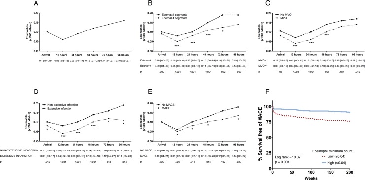
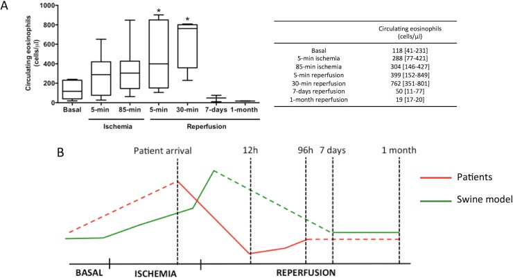
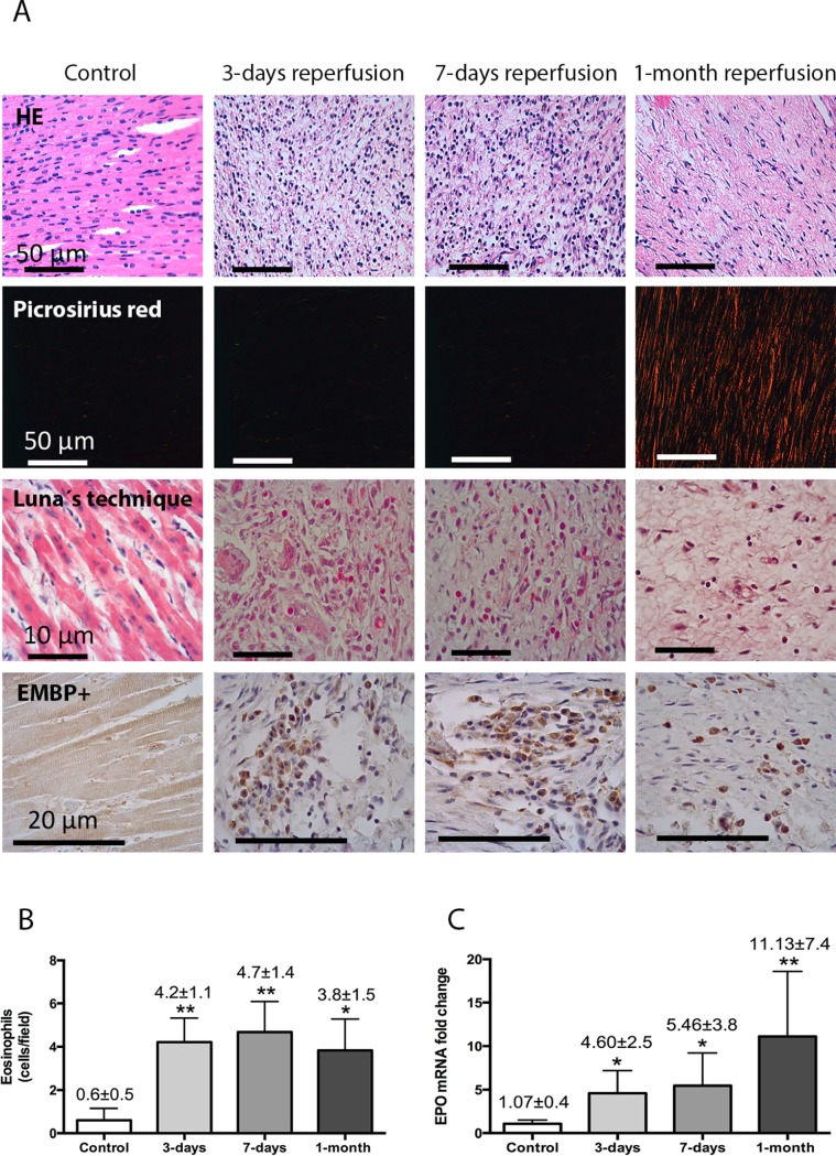
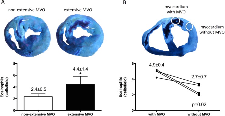
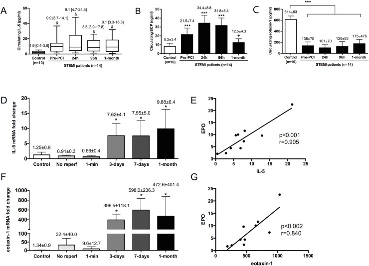
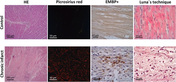
Results: Patient eosinophil count sharply decreased 12h post-reperfusion compared to arrival. A lower minimum eosinophil count was associated with more extensive edema, microvascular obstruction, and infarct size as measured by cardiac magnetic resonance, and also with a higher rate of cardiac events (death, re-infarction, or heart failure) during follow-up. In the experimental model, eosinophil count boosted during ischemia and dropped back immediately post-reperfusion. Myocardial samples revealed progressive eosinophil migration into the infarcted myocardium, especially areas with microvascular obstruction. Markers of eosinophil maturation and survival (interleukin-5), degranulation (eosinophil cationic protein) and migration (eotoxin-1) were detected in the blood of patients, and in porcine myocardium. Eosinophil infiltration was detected in autopsies from chronic MI patients.

Conclusion: Eosinopenia post-MI was associated with an impaired cardiac structure and adverse events. The decay in circulating eosinophils soon after reperfusion mirrors their migration into the infarcted myocardium, as reflected by their presence in heart samples from swine and patients. Further studies are needed to understanding this unexplored pathway and its therapeutic implications.


Conflict of interest statement

The authors have declared no competing interests exist.

Figures

Fig 1
Fig 1. Temporal evolution of eosinophils after reperfused ST-segment elevation myocardial infarction and in relation to edema, microvascular obstruction (MVO), infarct size, and major adverse cardiac events (MACE).
Temporal evolution of eosinophils (x1000 cells/ml) (A) in the whole study group and in relation to (B) edema, (C) MVO, (D) infarct size and (E) MACE (cardiac death, re-infarction, or readmission for heart failure) during follow-up. Data are expressed as median with the interquartile range (n = 620) and were analysed by Mann-Whitney U-test. (F) Eosinophil minimum count and outcome Kaplan-Meier curves for survival free of MACE (cardiac death, re-infarction, and readmission for heart failure) during follow-up depending on low eosinophil count. Abbreviations: LV: left ventricular.
Fig 2
Fig 2. Dynamics of circulating eosinophils in swine subjected to reperfused myocardial infarction (MI).
Whole blood was isolated from the experimental model at basal and at different time points of the ischemia and reperfusion process: after 5-min and 85-min of ischemia (immediately before reperfusion) as well as 5-minutes, 30-minutes, 7-days, and 1-month after reperfusion post-MI. Samples were incubated with FITC-CD45 and PE-CD16 and afterwards measured using flow cytometry. (A) Eosinophil counts in whole blood were analysed. Data were expressed as median with the interquartile range (n≥5 independent experiments) and were analysed by Kruskal-Wallis analysis followed by Dunn’s test. *P<0.05 vs. basal. (B) Dynamics of peripheral eosinophils in ST-elevation MI-patients (red) and in the swine model (green). The drop in peripheral eosinophils detected in ST-elevation MI-patients 12h after coronary reperfusion might be preceded by their progressive increase during ischemia and soon after reperfusion, as observed in the experimental model.
Fig 3
Fig 3. Eosinophil mobilization into the infarcted myocardium in a controlled swine model of reperfused myocardial infarction (MI).
(A) Representative images from infarcted tissue isolated from control and three MI groups (90-min of ischemia followed by 3-days, 7-days, and 1-month reperfusion) stained with hematoxylin-eosin (HE) (first panel). The presence of eosinophils was revealed by staining myocardial samples with Luna’s technique, specific for eosinophil granules (second panel) and with the eosinophil-specific protein eosinophil major basic protein (EMBP) (third panel). (B) Quantification of eosinophil cells in the myocardial tissue. Images from the infarcted area isolated from the four independent groups were quantified with Image-Pro Plus analysis software. Scoring was performed by a blinded observer unaware of the experimental group. (C) The expression of eosinophil peroxidase (EPO) in the infarcted myocardium at different times of the ischemia and reperfusion process. Data (mean±SD, n≥4) were analysed by one-way ANOVA analysis followed by Bonferroni test. *P<0.05, **P<0.01 vs. control.
Fig 4
Fig 4. Infiltration of eosinophils into the infarcted areas with microvascular obstruction (MVO).
(A) Quantification of infiltrated eosinophils according to the extension of MVO. Animals from the 3-days and 7-days reperfusion groups were categorized according to MVO (extensive: MVO> 5% of area at risk, median value) and the number of eosinophils was morphometrically quantified. (B) Quantification of infiltrated eosinophils in samples obtained from the same heart but comparing infarcted areas with macroscopic MVO and without MVO from the same heart.
Fig 5
Fig 5. Evolution of crucial cytokines implicated in eosinophil maturation, activation, and recruitment.
The serum levels of interleukin (IL)-5 (A), eosinophil cationic protein (ECP) (B), and eotaxin-1 (C) were determined in samples isolated from control subjects (n = 10) and from ST-elevation myocardial infarction (STEMI)-patients (n = 14) upon arrival and after primary coronary intervention (PCI) (24h, 96h, and 1-month). The expression in the infarcted myocardium of IL-5 (D), and eotaxin-1 (F) were obtained at different times of the ischemia and reperfusion process. The correlation of IL-5 and eotoxin-1 with the expression of eosinophil peroxidase (EPO) (E,G, respectively) was assessed using Pearson correlation coefficient. Continuous normally distributed data are expressed as mean±SD and were analysed by one-way ANOVA analysis followed by Bonferroni test. *P<0.05, ***P<0.001 vs. control; +P<0.05 vs. 24h post-PCI. Non-parametric data were expressed as the median with the interquartile range and were analysed by Kruskal-Wallis followed by Dunn’s test. &P<0.05 vs. control. Abbreviations: EMBP: eosinophil major basic protein.
Fig 6
Fig 6. The presence of eosinophils in autopsies from patients with chronic myocardial infarction (MI).
Representative images from infarcted tissue isolated from control and chronic MI patients stained with hematoxylin-eosin (HE) (left panel), and picrosirius red (middle-left panel). The presence of eosinophils was revealed by staining myocardial samples with the eosinophil-specific protein eosinophil major basic protein (EMBP) (middle-right panel) and with Luna’s technique, specific for eosinophil granules (right panel).

Similar articles

Cited by

References

    1. Blanchard C, Rothenberg ME. Biology of eosinophils. Adv Immunol. 2009;101: 81–121. 10.1016/S0065-2776(08)01003-1 - DOI - PMC - PubMed
    1. Davoine F, Lacy P. Eosinophil cytokines, chemokines, and growth factors: emerging roles in immunity. Front Immunol. 2014;5: 570 10.3389/fimmu.2014.00570 - DOI - PMC - PubMed
    1. Possa SS, Leick EA, Prado CM, Martins MA, Tiberio IF. Eosinophilic inflammation in allergic asthma. Front Pharmacol. 2013;4: 46 10.3389/fphar.2013.00046 - DOI - PMC - PubMed
    1. Diny NL, Hou X, Barin JG, Chen G, Talor MV, Schaub J, et al. Macrophages and cardiac fibroblasts are the main producers of eotaxins and regulate eosinophil trafficking to heart. Eur J Immunol. 2016;46: 2749–2760. 10.1002/eji.201646557 - DOI - PMC - PubMed
    1. Husser O, Bodi V, Sanchis J, Nuñez J, Mainar L, Chorro FJ, et al. White blood cell subtypes after STEMI: temporal evolution, association with cardiac magnetic resonance–derived infarct size and impact on outcome. Inflammation. 2011;34: 73–84. 10.1007/s10753-010-9209-0 - DOI - PubMed

Publication types