. 2018 Oct 26;14(10):e1007359.
doi: 10.1371/journal.ppat.1007359. eCollection 2018 Oct.

Integrin but not CEACAM receptors are dispensable for Helicobacter pylori CagA translocation

Affiliations

Integrin but not CEACAM receptors are dispensable for Helicobacter pylori CagA translocation

Qing Zhao et al. PLoS Pathog. .

Abstract

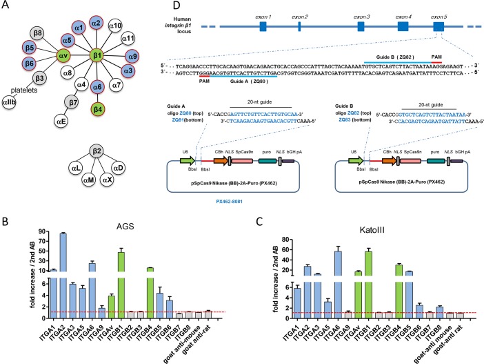
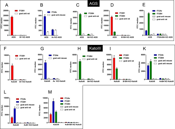
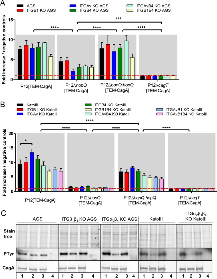
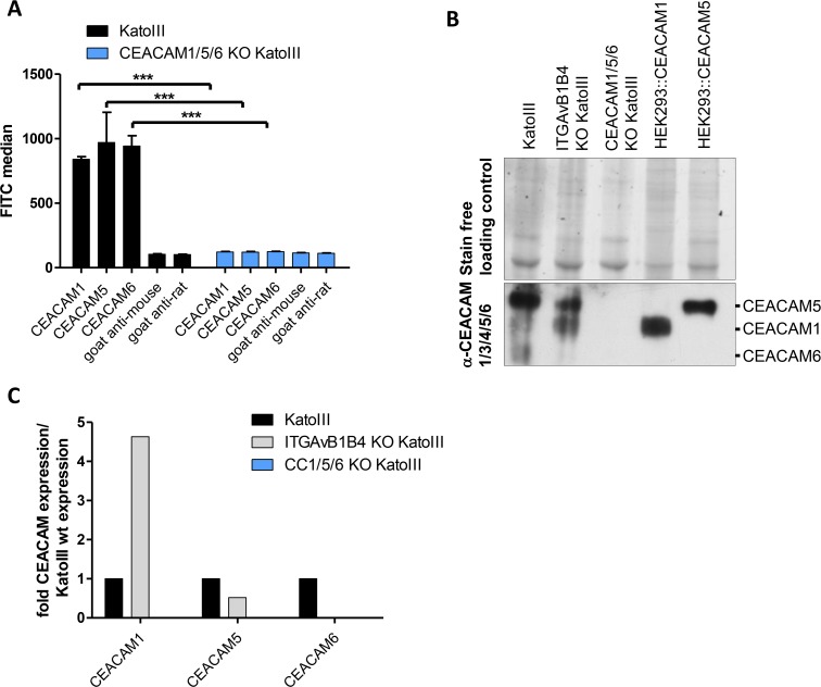
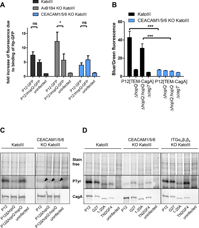
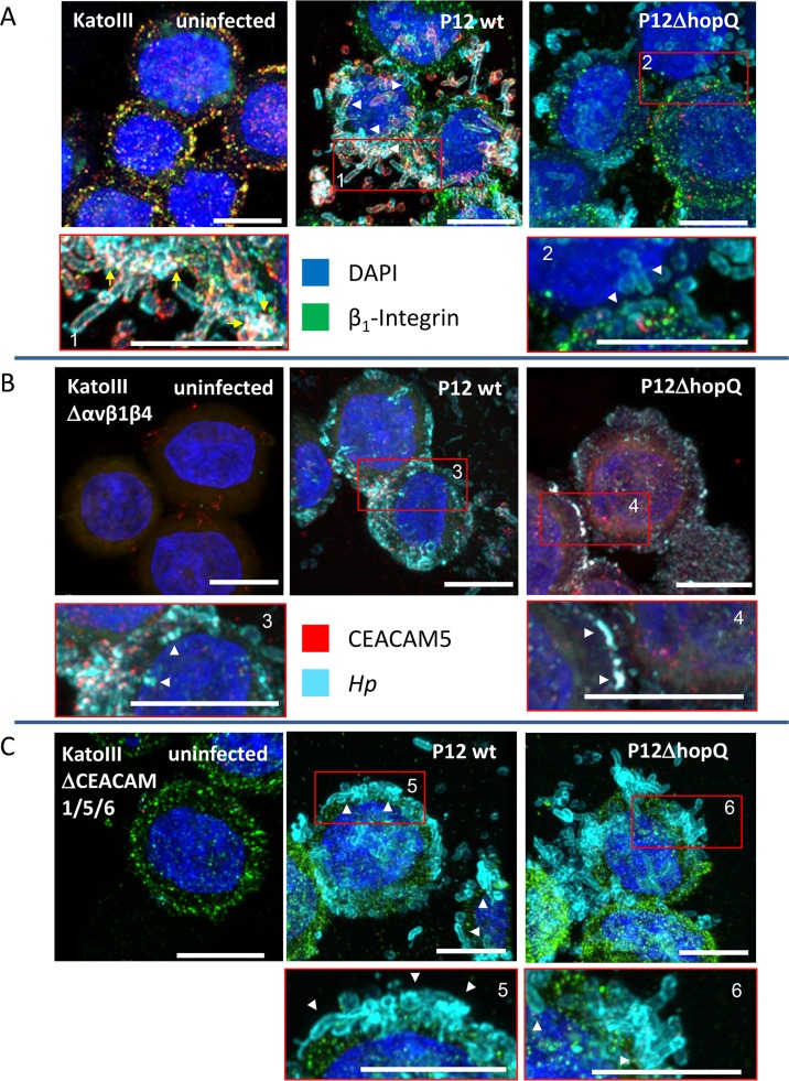
Translocation of the Helicobacter pylori (Hp) cytotoxin-associated gene A (CagA) effector protein via the cag-Type IV Secretion System (cag-T4SS) into host cells is a hallmark of infection with Hp and a major risk factor for severe gastric diseases, including gastric cancer. To mediate the injection of CagA, Hp uses a membrane-embedded syringe-like molecular apparatus extended by an external pilus-like rod structure that binds host cell surface integrin heterodimers. It is still largely unclear how the interaction of the cag-T4SS finally mediates translocation of the CagA protein into the cell cytoplasm. Recently certain carcinoembryonic antigen-related cell adhesion molecules (CEACAMs), acting as receptor for the Hp outer membrane adhesin HopQ, have been identified to be involved in the process of CagA host cell injection. Here, we applied the CRISPR/Cas9-knockout technology to generate defined human gastric AGS and KatoIII integrin knockout cell lines. Although confocal laser scanning microscopy revealed a co-localization of Hp and β1 integrin heterodimers on gastric epithelial cells, Hp infection studies using the quantitative and highly sensitive Hp β-lactamase reporter system clearly show that neither β1 integrin heterodimers (α1β1, α2β1 or α5β1), nor any other αβ integrin heterodimers on the cell surface are essential for CagA translocation. In contrast, deletion of the HopQ adhesin in Hp, or the simultaneous knockout of the receptors CEACAM1, CEACAM5 and CEACAM6 in KatoIII cells abolished CagA injection nearly completely, although bacterial binding was only reduced to 50%. These data provide genetic evidence that the cag-T4SS-mediated interaction of Hp with cell surface integrins on human gastric epithelial cells is not essential for CagA translocation, but interaction of Hp with CEACAM receptors is facilitating CagA translocation by the cag-T4SS of this important microbe.


Conflict of interest statement

The authors have declared that no competing interests exist.

Figures

Fig 1
Fig 1. Schematic representation of the mammalian integrin receptor family, integrin profiling in AGS and KatoIII gastric cell lines and the strategy for integrin β1 knockout generation.
A) Illustration of possible integrin α and β associations [21]. Epithelial cell-specific heterodimers are marked with red circles, α and β subunits expressed in AGS or KatoIII cells, as determined in B and C, are shown as filled blue or green (integrin genes targeted by CRISPR/Cas-mediated gene knockout) circles. Grey and white circles represent subunits tested but not expressed, or not tested for expression, respectively. B) Integrin expression profile of AGS cells as determined by flow cytometry using different integrin antibodies. C) Integrin expression profile of KatoIII cells as determined by flow cytometry. D) Strategy for targeted deletion of integrin β1 gene. Streptococcus pyogenes Cas9 nickase binding sites (20 bp, highlighted in blue) are immediately followed by the 5’-NGG PAM (protospacer adjacent motif). The short guide RNA (sgRNA) pairs are located on both strands of the target DNA with a 25 bp gap. Cloning scheme of the CRISPR plasmids (see Materials and methods for details). All values in B and C were determined as standard errors of the mean (±SEM) from three independent experiments.
Fig 2
Fig 2. Integrin expression of AGS and KatoIII wild type and corresponding single and multiple integrin-knockout cell lines.
A-E) Integrin expression was determined showing FITC median from three independent flow cytometry experiments. As negative controls, cells were stained with secondary antibody only (Goat-anti mouse, Goat-anti rat). A) ITGB1 surface expression in wild type and ITGB1 KO AGS cells. B) ITGAv surface expression in wild type and ITGAv KO AGS cells. C) ITGB4 surface expression in wild type and ITGB4 KO AGS cells. D) ITGB1 and ITGB4 surface expression in wild type and ITGB1B4 KO AGS cells. E) ITGAv and ITGB4 surface expression in wild type and ITGAvB4 KO AGS cells. F-M) Integrin expression was determined showing FITC median from three independent flow cytometry experiments. As negative controls, cells were stained with secondary antibody only (Goat-anti mouse, Goat-anti rat). F) ITGB1 surface expression in wild type and ITGB1 KO KatoIII cells. G) ITGAv surface expression in wild type and ITGAv KO KatoIII cells. H) ITGB4 surface expression in wild type and ITGB4 KO KatoIII cells. I) ITGAv and ITGB1 surface expression in wild type and ITGAvB1 KO KatoIII cells. K) ITGAv and ITGB4 surface expression in wild type and ITGAvB4 KO KatoIII cells. L) ITGB1 and ITGB4 surface expression in wild type and ITGB1B4 KO KatoIII cells. M) ITGAv, ITGB1 and ITGB4 surface expression in wild type and ITGB1AvB4 KO KatoIII cells. All values are indicated as average values including standard errors of the mean (±SEM), (n = 3).
Fig 3
Fig 3. CagA tyrosine phosphorylation and quantitative evaluation of CagA translocation into wild type integrin-knockout AGS or KatoIII cell lines by the TEM-1 β-lactamase reporter assay.
A) AGS and five integrin-depletion cell lines in 96-well plates were infected with P12[TEM-CagA], the P12ΔhopQ[TEM-CagA], the genetically complemented P12ΔhopQ:hopQ[TEM-CagA] and as negative control the translocation-deficient P12ΔcagT[TEM-CagA] deletion mutant at an MOI of 60. B) KatoIII wild type and seven single or multiple integrin depletion cell lines in 96-well plates were infected with P12[TEM-CagA], the P12ΔhopQ[TEM-CagA], the genetically complemented P12ΔhopQ:hopQ[TEM-CagA] and as negative control the translocation-deficient P12ΔcagT[TEM-CagA] deletion mutant at an MOI of 60. Ratios of blue to green fluorescence of each sample were calculated and normalized to the mean of blue to green ratio of the negative controls. All values were indicated as standard errors of the mean (±SEM) from n = 3 independent experiment for AGS and n = 5 independent experiments for KatoIII cells. The red line marks the level of the controls. C) AGS cells, two AGS integrin-depletion cell lines (ITGB1B4 KO, ITGAvB4 KO), KatoIII cells and the triple integrin-depletion KatoIII cell line (ITGAvB1B4 KO) were infected with strain P12, P12ΔhopQ or P12ΔhopQ:hopQ, for 2.5 hours with an MOI of 60. Translocation of CagA was determined by detecting tyrosine-phosphorylated CagA with the anti phosphotyrosine antibody PY99. Statistics: Two-way ANOVA with a Tukey’s multiple comparison Post-Hoc test was performed. (* P < 0.05; ** P < 0.01, *** P < 0.001, **** P < 0.0001). Values within each group did not change significantly, with exception of KatoIII wild type versus ITGAv KO KatoIII cells infected by P12 [TEM-CagA].
Fig 4
Fig 4. Characterization of KatoIII wild type and CEACAM- or integrin knockout cells for CEACAM expression by flow cytometry and western blotting.
A) Wild type and CEACAM1/5/6 KO KatoIII cells were analyzed for CEACAM receptors on their surface by flow cytometry using anti-CEACAM antibodies (CEACAM1 (8G5, Genovac), CEACAM5 (26/3/13, Genovac), CEACAM6 (9A6, Genovac)). Goat anti-mouse or anti-rat were used as negative control antibodies (n = 3). For statistical analysis the Two-way ANOVA with Tukey’s HSD post-test was performed. (ns, non-significant, *** p<0.001). B) Immunoblot showing the production of CEACAM1, CEACAM5 and CEACAM6 by KatoIII cells and the absence of the receptors in KatoIII CEACAM1/5/6 KO cells, as indicated. The Pan α-CEACAM antibody (CEACAM1/3/4/5/6 (D14HD11, Genovac)), recognizing all three CEACAMs was used. Shown is a representative blot of an experiment that was performed three times. Lysates of HEK293::CEACAM1 and HEK293::CEACAM5 producing cells were added as controls for the correct size of the corresponding CEACAM proteins. The stainfree method was used as loading control. C) Densitometric quantification of CEACAM band signals in each sample in the immunoblot presented as fold CEACAM expression compared to Kato wild type cells.
Fig 5
Fig 5. KatoIII wild type, KatoIIIΔανβ1β4 and KatoIII CEACAM1/5/6 KO cells tested for binding of P12 wt and P12ΔhopQ mutant strains and their CagA translocation capacity.
A) P12-GFP and P12ΔhopQ-GFP strains were used for infection of KatoIII wild type, KatoIIIΔανβ1β4 and KatoIII CEACAM1/5/6 KO cells. The bacterial binding capacity of Hp P12-GFP and a P12ΔhopQ-GFP strain to the different cell lines was evaluated by flow cytometry (n = 4). The data are normalized to uninfected KatoIII cells. Statistics: Data were analyzed by Two-way ANOVA. As Post-Hoc Test a Tukey’s multiple comparison test was performed. (ns: not significant; * P < 0.05). B) KatoIII wild type and KatoIII CEACAM1/5/6 KO cells were infected with Hp P12[TEM-CagA] and corresponding mutant strains at an MOI of 60 for 2.5 h, as indicated. Ratios of blue to green fluorescence of each sample were calculated and normalized to the mean of blue to green ratio of the negative controls. All values were indicated as standard errors of the mean (±SEM) from n = 5 independent experiments. Statistics: Two-way ANOVA was performed. As Post-Hoc test mutants mean were compared by a Bonferroni test (ns: not significant *** P < 0.001). C) KatoIII cells or the CEACAM1/5/6 KO cell line were infected with strain P12, P12ΔhopQ or P12ΔhopQ:hopQ, for 2.5 hours with an MOI of 60. Translocation of CagA was determined by detecting tyrosine-phosphorylated CagA with the antibody PY99. Arrowheads indicate the position of the weak tyrosine-phosphorylated (PTyr) CagA band. D) KatoIII cells or the triple integrin-depletion KatoIII cell line were infected with strain P12, G27, 1-20A or TN2GF4 for 2.5 h with an MOI of 60. Translocation of CagA was determined by detecting tyrosine-phosphorylated CagA with the antibody PY99.
Fig 6
Fig 6. Confocal laser scanning microscopy (CLSM) studies of the epithelial cell line KatoIII and integrin and CEACAM knockout KatoIII cells infected with Hp P12 wild type and mutant strains.
Confluent monolayers of KatoIII (A), KatoIIIΔαvβ1β4 (B) and or KatoIIIΔCEACAM1/5/6 (C) cells were infected with Hp P12 wt, P12ΔhopQ or P12ΔhopQ::hopQI. The cells were analyzed by CLSM (63 x objective) by a Zeiss LSM 880 with Airyscan. The top row shows an overview, the lower row a magnification of characteristic observation fields specified by a red window. Several planes are combined to obtain a Z-stack (bottom to top), showing bacterial binding and co-localization of β1 integrin or CEACAM5 and Hp. White arrowheads point to the close association of Hp with the gastric epithelial cell surface, yellow arrows point to co-localization events between Hp and CEACAM. Scale bars represent 10 μm. At least 3 micrographs of independent cell culture samples were taken, one representative area is shown. Colors for co-localizations: β1-Integrin/CEACAM5 (green/red): yellow; CEACAM5/ Hp (red/petrol-light blue): white; β1-Integrin/CEACAM5/Hp (green/red, petrol-light blue): white.

Comment in

Similar articles

Cited by

References

    1. Costa TR, Felisberto-Rodrigues C, Meir A, Prevost MS, Redzej A, Trokter M, et al. Secretion systems in Gram-negative bacteria: structural and mechanistic insights. Nat Rev Microbiol. 2015;13(6):343–59. nrmicro3456 [pii]; 10.1038/nrmicro3456 - DOI - PubMed
    1. Fronzes R, Christie PJ, Waksman G. The structural biology of type IV secretion systems. Nat Rev Microbiol. 2009;7(10):703–14. nrmicro2218 [pii]; 10.1038/nrmicro2218 - DOI - PMC - PubMed
    1. Alvarez-Martinez CE, Christie PJ. Biological diversity of prokaryotic type IV secretion systems. Microbiol Mol Biol Rev. 2009;73(4):775–808. 73/4/775 [pii]; 10.1128/MMBR.00023-09 - DOI - PMC - PubMed
    1. Censini S, Lange C, Xiang ZY, Crabtree JE, Ghiara P, Borodovsky M, et al. Cag, a pathogenicity island of Helicobacter pylori, encodes type I-specific and disease-associated virulence factors. Proc Natl Acad Sci USA. 1996;93:14648–53. - PMC - PubMed
    1. Odenbreit S, Püls J, Sedlmaier B, Gerland E, Fischer W, Haas R. Translocation of Helicobacter pylori CagA into gastric epithelial cells by type IV secretion. Science. 2000;287:1497–500. - PubMed

Publication types

MeSH terms