. 2018 Oct 26;13(10):e0206104.
doi: 10.1371/journal.pone.0206104. eCollection 2018.

Superresolution microscopy with novel BODIPY-based fluorophores

Affiliations

Superresolution microscopy with novel BODIPY-based fluorophores

Amy M Bittel et al. PLoS One. .

Abstract

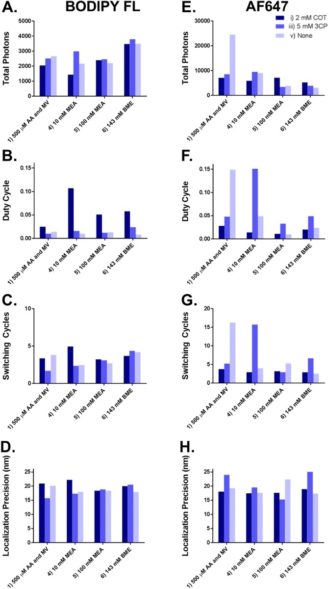
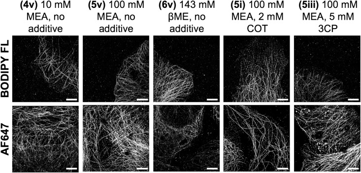
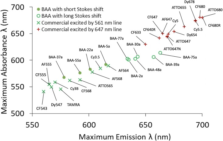
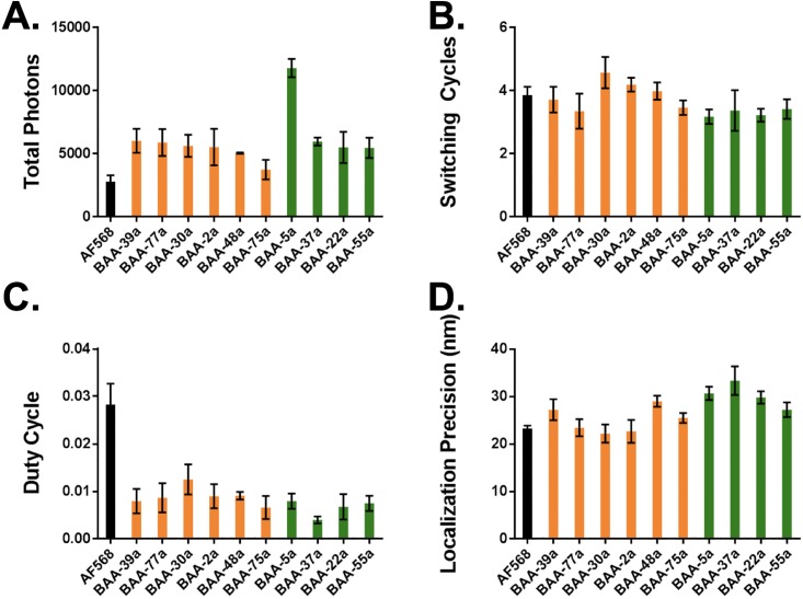
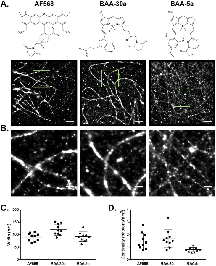
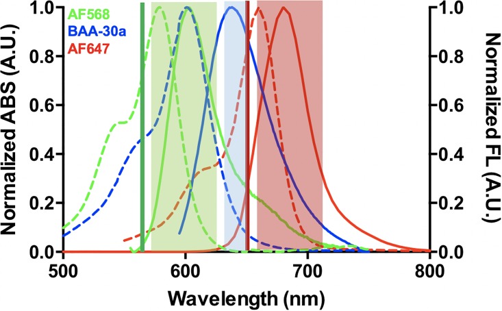
Multicolor single-molecule localization microscopy (SMLM) expands our understanding of subcellular details and enables the study of biomolecular interactions through precise visualization of multiple molecules in a single sample with resolution of ~10-20 nm. Probe selection is vital to multicolor SMLM, as the fluorophores must not only exhibit minimal spectral crosstalk, but also be compatible with the same photochemical conditions that promote fluorophore photoswitching. While there are numerous commercially available photoswitchable fluorophores that are optimally excited in the standard Cy3 channel, they are restricted to short Stokes shifts (<30 nm), limiting the number of colors that can be resolved in a single sample. Furthermore, while imaging buffers have been thoroughly examined for commonly used fluorophore scaffolds including cyanine, rhodamine, and oxazine, optimal conditions have not been found for the BODIPY scaffold, precluding its routine use for multicolor SMLM. Herein, we screened common imaging buffer conditions including seven redox reagents with five additives, resulting in 35 overall imaging buffer conditions to identify compatible combinations for BODIPY-based fluorophores. We then demonstrated that novel, photoswitchable BODIPY-based fluorophores with varied length Stokes shifts provide additional color options for SMLM using a combination of BODIPY-based and commercially available photoswitchable fluorophores.


Conflict of interest statement

Dr. Nick Dolman is an employee of Thermo Fisher Scientific. Participation in this work does not alter our adherence to PLOS ONE policies on sharing data and materials.

Figures

Fig 1
Fig 1. Photoswitching property quantification.
The photoswitching properties of single fluorophore molecules isolated in PVA including total photon output, duty cycle, number of switching cycles and localization precision were quantified for (A)–(D) BODIPY FL and (E)–(G) AF647. The photoswitching properties were quantified in (1) 500 μM AA and MV, (4) 10 mM MEA, (5) 100 mM MEA and (6) 143 mM βME with the additives (i) 2 mM COT, (iii) 5 mM 3CP, and (v) no additive, results in 12 image buffer conditions tested per fluorophore.
Fig 2
Fig 2. SMLM imaging of immunolabeled microtubules using a subset of imaging buffer conditions.
SMLM imaging of immunolabeled microtubules in vitro resulted in varied image quality using BODIPY FL (top) and AF647 (bottom). Scale bar = 5 μm.
Fig 3
Fig 3. Conventional fluorophore spectral emission space gap.
The maximum absorbance wavelength vs. maximum emission wavelength for conventional fluorophores excited using a standard Cy3 imaging channel/561 nm laser line (x) and Cy5 imaging channel/647 nm laser line (+) result in a gap in spectral emission space from 615–650 nm. This spectral space can be filled using novel BAA fluorophores with long Stokes shifts (○) [13]. Four additional BAA fluorophores with short Stokes shifts (●) were also evaluated for their SMLM imaging utility. Each point is labeled with the fluorophore name.
Fig 4
Fig 4. Photoswitching properties for long and short Stokes shift BAA fluorophores.
Photoswitching properties including (A) total photons, (B) number of switching cycles, (C) duty cycle and (D) localization precision were quantified for AF568 (black), six BAA fluorophores with long Stokes shifts (orange), and four BAA fluorophores with short Stokes shifts (green) [13]. The average +/- standard deviation of triplicate measurements of single molecules isolated in PVA are reported.
Fig 5
Fig 5. SMLM imaging with long and short Stokes shift BAA fluorophores.
(A) SMLM imaging of immunolabeled microtubules in vitro via indirect immunofluorescence in fixed cells labeled with AF568, BAA-30a and BAA-5a, where the chemical structures of each fluorophore containing its conjugatable NHS ester group is shown. Scale bar = 2 μm. (B) Magnified images to show further detail of the boxed regions of (A) Scale bar = 0.5 μm. The average microtubule (C) width and (D) continuity for each fluorophore were calculated and are shown as the mean ± 25% of n = 10 measurements per image of the corresponding fluorophore.
Fig 6
Fig 6. Potential optical configuration for 3-color SMLM.
The normalized absorbance (dotted, right axis) and emission (solid, left axis) spectra of AF568, long Stokes shift BAA fluorophore BAA-30a and AF647 are shown. The 561 nm (green) and 647 nm (red) laser lines are shown with transmission of conventional Cy3 (green, 605/70 nm) and Cy5 (red, 700/75 nm) bandpass filters as well as an additional bandpass filter optimal for BAA-30a detection (blue, 640/20 nm) permitting detection of all three fluorophores with minimal cross talk for SMLM.

Similar articles

Cited by

References

    1. Betzig E, Patterson GH, Sougrat R, Lindwasser OW, Olenych S, Bonifacino JS, et al. Imaging intracellular fluorescent proteins at nanometer resolution. Science. 2006;313(5793):1642–5. 10.1126/science.1127344 . - DOI - PubMed
    1. Rust MJ, Bates M, Zhuang X. Sub-diffraction-limit imaging by stochastic optical reconstruction microscopy (STORM). Nature methods. 2006;3(10):793–5. 10.1038/nmeth929 . - DOI - PMC - PubMed
    1. Xu K, Babcock HP, Zhuang X. Dual-objective STORM reveals three-dimensional filament organization in the actin cytoskeleton. Nature methods. 2012;9(2):185–8. Epub 2012/01/11. 10.1038/nmeth.1841 . - DOI - PMC - PubMed
    1. Heilemann M, van de Linde S, Schuttpelz M, Kasper R, Seefeldt B, Mukherjee A, et al. Subdiffraction-resolution fluorescence imaging with conventional fluorescent probes. Angew Chem Int Ed Engl. 2008;47(33):6172–6. Epub 2008/07/23. 10.1002/anie.200802376 . - DOI - PubMed
    1. van de Linde S, Loschberger A, Klein T, Heidbreder M, Wolter S, Heilemann M, et al. Direct stochastic optical reconstruction microscopy with standard fluorescent probes. Nature protocols. 2011;6(7):991–1009. 10.1038/nprot.2011.336 . - DOI - PubMed

Publication types