. 2018 Oct 25;10(11):582.
doi: 10.3390/v10110582.

Evidence of Human Parvovirus B19 Infection in the Post-Mortem Brain Tissue of the Elderly

Affiliations

Evidence of Human Parvovirus B19 Infection in the Post-Mortem Brain Tissue of the Elderly

Sandra Skuja et al. Viruses. .

Abstract

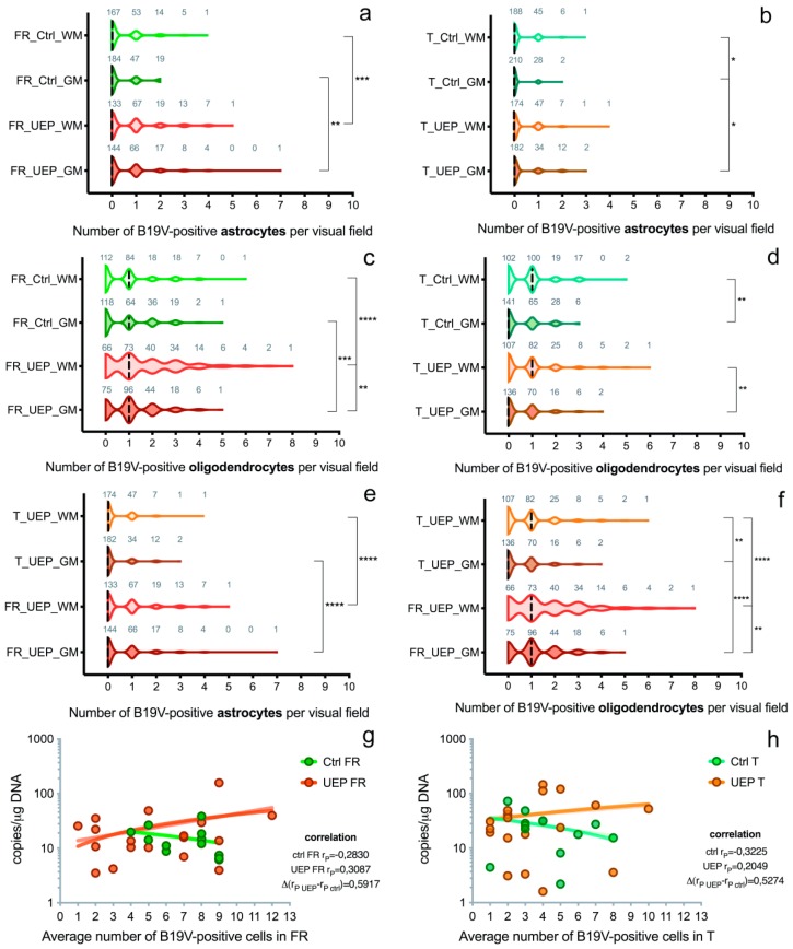
After primary exposure, the human parvovirus B19 (B19V) genome may remain in the central nervous system (CNS), establishing a lifelong latency. The structural characteristics and functions of the infected cells are essential for the virus to complete its life cycle. Although B19V has been detected in the brain tissue by sequencing PCR products, little is known about its in vivo cell tropism and pathogenic potential in the CNS. To detect B19V and investigate the distribution of its target cells in the CNS, we studied brain autopsies of elderly subjects using molecular virology, and optical and electron microscopy methods. Our study detected B19V in brain tissue samples from both encephalopathy and control groups, suggesting virus persistence within the CNS throughout the host's lifetime. It appears that within the CNS, the main target of B19V is oligodendrocytes. The greatest number of B19V-positive oligodendrocytes was found in the white matter of the frontal lobe. The number was significantly lower in the gray matter of the frontal lobe (p = 0.008) and the gray and white matter of the temporal lobes (p < 0.0001). The morphological changes observed in the encephalopathy group, propose a possible B19V involvement in the demyelination process.

Keywords: PCR; elderly; electron microscopy; glia; immunohistochemistry; parvovirus B19.


Conflict of interest statement

The authors declare no conflict of interest.

Figures

Figure 1
Figure 1
Representation of data from the brain tissue analysis: (a) Copies of B19V VP1 genomic sequence in the frontal lobes of control (Ctrl) and unspecified encephalopathy (UEP) groups; (b) Copies of B19V VP1 genomic sequence in the temporal lobes of control (Ctrl) and UEP groups. Scatter plots with bar represent geometric mean ± 95% CI.
Figure 2
Figure 2
Data representing brain tissue analysis: (a) Distribution of B19V-positive astrocytes per visual fields in the gray (GM) and white matter (WM) of the UEP group and controls (Ctrl), frontal lobe (FR); (b) Distribution of B19V-positive astrocytes per visual fields in the gray (GM) and white matter (WM) of the UEP group and controls (Ctrl), temporal lobe (T); (c) Distribution of B19V-positive oligodendrocytes per visual fields in the gray (GM) and white matter (WM) of the UEP group and controls (Ctrl), frontal lobe (FR); (d) Distribution of B19V-positive oligodendrocytes per visual fields in the gray (GM) and white matter (WM) of the UEP group and controls (Ctrl), temporal lobe (T); (e) Distribution of B19V-positive astrocytes per visual fields in the gray (GM) and white matter (WM) of the UEP group, frontal lobe (FR), temporal lobe (T); (f) Distribution of B19V-positive oligodendrocytes per visual fields in the gray (GM) and white matter (WM) of the UEP group, frontal lobe (FR), temporal lobe (T); (g) Correlation of qPCR (y-axis) and IHC (x-axis) data in the frontal lobe (FR) tissue samples, comparison of regression slopes of the control and UEP groups; (h) Correlation of qPCR (y-axis) and IHC (x-axis) data in the temporal lobe (T) tissue samples, comparison of regression slopes of the control and UEP groups. Violin plots: dashed lines represent the approximate ratio of visual fields (out of 240) with B19V-positive cells to fields with B19V-negative cells (“0”—ratio less than 1.0, “1”—more than 1.0); numbers in gray show visual fields; asterisks represent a significance level (* p < 0.05, ** p < 0.01, *** p < 0.001, **** p < 0.0001) of differences between groups (Kruskal–Wallis test followed by two-stage step-up method of Benjamini, Krieger, and Yekutieli as post hoc procedure).
Figure 3
Figure 3
IHC: (a) B19V-positive oligodendrocytes (black arrows) and endotheliocytes (white arrowheads) in the gray matter of the UEP subject (frontal lobe, 400×); (b) B19V-positive oligodendrocytes (black arrows), astrocytes (white arrows) and endotheliocyte (white arrowhead) in the white matter of the UEP subject (frontal lobe, 400×); (c) B19V-positive oligodendrocyte (black arrow) and astrocyte (white arrow) in the gray matter of the UEP subject (temporal lobe, 400×); (d) B19V-positive oligodendrocytes (black arrows), astrocytes (white arrows), endotheliocytes (white arrowhead) and erythrocytes (black arrowhead) in the white matter of the UEP subject (temporal lobe, 400×); (e) myelin basic protein (MBP) immunoexpression in the white matter of the UEP subject (frontal lobe, 400×); (f) MBP immunoexpression in the white matter of the UEP subject (temporal lobe, 400×).
Figure 4
Figure 4
Detection of viral capsid antigens by immunofluorescence, confocal microscopy, DAPI—blue, B19V-immunopositive products—green: (a) B19V-positive oligodendrocytes and neurons in the gray matter of the UEP subject (frontal lobe, 1000×), the insert shows magnified view of the oligodendrocyte; (b) B19V-positive oligodendrocytes in the white matter of the UEP subject (frontal lobe, 1000×); (c) B19V-positive oligodendrocytes in the gray matter of the UEP subject (temporal lobe, 1000×); (d) B19V-positive oligodendrocyte in the white matter of the UEP subject (temporal lobe, 1000×).
Figure 4
Figure 4
Detection of viral capsid antigens by immunofluorescence, confocal microscopy, DAPI—blue, B19V-immunopositive products—green: (a) B19V-positive oligodendrocytes and neurons in the gray matter of the UEP subject (frontal lobe, 1000×), the insert shows magnified view of the oligodendrocyte; (b) B19V-positive oligodendrocytes in the white matter of the UEP subject (frontal lobe, 1000×); (c) B19V-positive oligodendrocytes in the gray matter of the UEP subject (temporal lobe, 1000×); (d) B19V-positive oligodendrocyte in the white matter of the UEP subject (temporal lobe, 1000×).
Figure 5
Figure 5
Immunogold staining, TEM: (a) Immunolabeled neuronal soma demonstrating the presence of the viral proteins in the nucleus (small dots showed by arrows, 50,000×); (b) (small dots showed by arrows, 30,000×). Nuclei show electron dense (dark) heterochromatin and electron lucid (light) euchromatin enclosed by nuclear envelope.

Similar articles

Cited by

References

    1. Licastro F., Carbone I., Raschi E., Porcellini E. The 21st Century Epidemic: Infections as Inductors of Neuro-Degeneration Associated with Alzheimer’s Disease. Immun. Ageing. 2014;11:22. doi: 10.1186/s12979-014-0022-8. - DOI - PMC - PubMed
    1. Swanson P.A., McGavern D.B. Viral Diseases of the Central Nervous System. Curr. Opin. Virol. 2015;11:44–54. doi: 10.1016/j.coviro.2014.12.009. - DOI - PMC - PubMed
    1. Barah F., Whiteside S., Batista S., Morris J. Neurological Aspects of Human Parvovirus B19 Infection: A Systematic Review. Rev. Med. Virol. 2014;24:154–168. doi: 10.1002/rmv.1782. - DOI - PMC - PubMed
    1. Young N.S., Brown K.E. Parvovirus B19. N. Engl. J. Med. 2004;350:586–597. doi: 10.1056/NEJMra030840. - DOI - PubMed
    1. Hokynar K., Norja P., Hedman K., Söderlund-Venermo M. Tissue Persistence and Prevalence of B19 Virus Types 1-3. Future Virol. 2007;2:377–388. doi: 10.2217/17460794.2.4.377. - DOI

LinkOut - more resources