. 2018 Oct 25;10(11):1576.
doi: 10.3390/nu10111576.

Tart Cherry Reduces Inflammation in Adipose Tissue of Zucker Fatty Rats and Cultured 3T3-L1 Adipocytes

Affiliations

Tart Cherry Reduces Inflammation in Adipose Tissue of Zucker Fatty Rats and Cultured 3T3-L1 Adipocytes

Shasika Jayarathne et al. Nutrients. .

Abstract

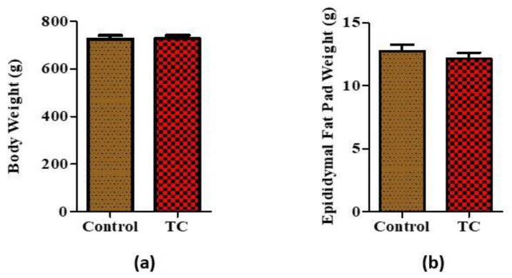
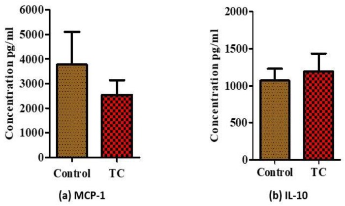
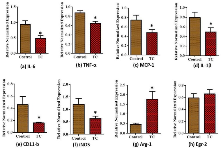
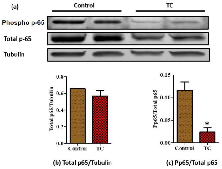
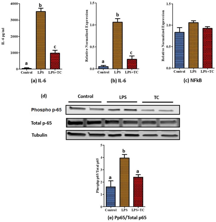
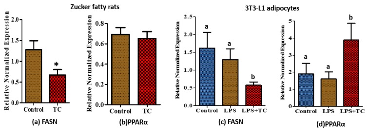
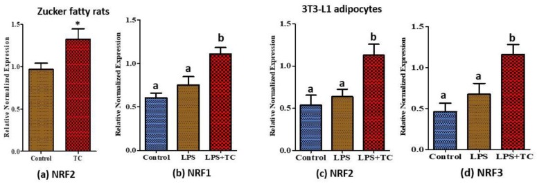
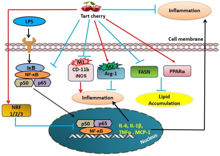
Obesity increases adipose tissue inflammation and secretion of pro-inflammatory adipokines, which have systemic effects on the organism's health status. Our objective was to dissect mechanisms of anti-inflammatory effects of tart cherry (TC) in adipose tissue of Zucker fatty rats, and cultured 3T3-L1 adipocytes. Rats were fed either a control diet, or 4% TC powder diets for eight weeks. Body and epididymal fat pad weights were not significantly different between control and TC groups. However, rats fed the TC diet had significantly reduced adipose tissue inflammation (p < 0.05), as determined by reduced mRNA levels of pro-inflammatory markers including interleukin-6 (IL-6), tumor necrosis factor alpha (TNFα), interleukin-1beta (IL-1β), monocyte chemoattractant protein 1 (MCP-1), inducible nitric oxide synthase (iNOS), and CD-11b, and increased mRNA levels of type-1 arginase (Arg-1) anti-inflammatory marker. Consistent with these in vivo results, TC significantly decreased expression of IL-6 mRNA and protein levels in lipopolysaccharide (LPS) stimulated adipocytes compared to those stimulated with LPS, but no TC. Moreover, both in vivo (rat adipose tissue) and in vitro (3T3-L1 adipocytes), phosphorylation of p65-NF-κB subunit was significantly reduced by TC. Additionally, TC decreased mRNA expression of fatty acid synthase (FASN), and increased expression of peroxisome proliferator-activated receptor alpha (PPARα), master regulator of lipid oxidation, and anti-oxidant markers nuclear factor erythroid-derived 2-related factor (NRFs) in both models. In conclusion, our findings indicate that TC downregulates inflammation in part via the nuclear factor kappa B (NF-κB) pathway in adipose tissue. Thus, TC may serve as a potential intervention to reduce obesity-associated inflammation.

Keywords: adipose tissue; inflammation; obesity; tart cherry.


Conflict of interest statement

S.J., A.M., S.S., J.H.K., K.C.-L. and N.M.-M. declare no conflict of interest. TC was a gift from the Cherry Marketing Institute to K.C.-L. and N.M.-M., A.J.S. received research funds from the Cherry Marketing Institute (CMI) for the animal study; however, CMI had no role or involvement in any part of this research.

Figures

Figure 1
Figure 1
Body weight and epididymal fat pad weight measured at week 8 between control and tart cherry (TC) fed group. (a) Body weight measurements at week 8, (b) epididymal fat pad weights. (n = 11/group).
Figure 2
Figure 2
Changes of serum inflammatory markers. (a) Serum levels of proinflammatory monocyte chemotactic protein 1 (MCP-1), and (b) serum levels of anti-inflammatory interleukin 10 (IL-10) in Zucker fatty rats. (n = 11/group).
Figure 3
Figure 3
Relative normalized mRNA expression of inflammatory markers in epididymal fat of Zucker fatty rats. (a) Interleukin-6 (IL-6), (b) tumor necrosis factor alpha (TNF-α), (c) monocyte chemoattractant protein-1 (MCP-1), (d) interleukin-1beta (IL-1β) pro-inflammatory markers, (e) CD11-b M1 marker, (f) inducible nitric oxide (iNOS) M1 marker, (g) type 1 arginase (Arg-1) M2 marker, and (h) early growth response protein 2 (Egr-2) M2 marker in epididymal adipose tissue of Zucker fatty rats. * p < 0.05, control vs. tart cherry (TC), (n > 9/group).
Figure 4
Figure 4
Expression levels of p-65 and tubulin proteins in epididymal fat of Zucker fatty rats detected by western blotting. (a) Phospho p-65, total p-65, and tubulin protein levels were examined using western blot. Tubulin was used as an internal loading control; (b) total p-65/Tubulin, (c) phospho p-65/Total p-65 (n = 8/group), two samples per group were used as representative images. * p < 0.05, control vs. tart cherry (TC).
Figure 5
Figure 5
Cell viability of 3T3-L1 cells using different concentrations of tart cherry (TC) extract. MTT assay was performed to assess potential toxic effects of different concentrations of TC extracts. Tart cherry has no toxic effects on 3T3-L1 adipocytes, two independent experiments.
Figure 6
Figure 6
Inflammatory protein and relative normalized mRNA expression in tart cherry (TC) treated 3T3-L1 adipocytes. Cells were pre-treated for 4 h with 12 µL/mL TC extract, then media was changed to 200 ng/mL lipopolysaccharide (LPS) with or without TC while the control had normal media. (a) IL-6 secretion from 3T3-L1 adipocytes into media, (b) IL-6 mRNA expression, and (c) nuclear factor kappa B (NF-κB) mRNA expression, (d) phosphor-p-65, total p-65, and tubulin protein levels were examined using western blot. For measuring the expression of phospho protein, cells were pretreated for 21 h with 12 µL/mL TC extract, then media was changed to 200 ng/mL LPS with or without TC while the control had normal media. Tubulin was used as an internal loading control, (e) Phospho p-65/Total p-65. a, b, c different letters are significantly different, p < 0.05, four independent experiments.
Figure 7
Figure 7
Relative normalized mRNA expression of fatty acid metabolic genes regulated by tart cherry (TC). (a) Expression of fatty acid synthase (FASN), (b) peroxisome proliferator-activated receptor alpha (PPARα) in epididymal adipose tissue of TC fed rats. * p < 0.05, control vs. tart cherry (TC), (n = 11/group), and (c) expression of FASN, (d) PPARα in TC treated 3T3-L1 adipocytes. a, b different letters are significantly different, p < 0.05, three independent experiments.
Figure 8
Figure 8
Relative normalized mRNA expression of anti-oxidant genes regulated by tart cherry (TC). (a) Expression of nuclear factor erythroid-derived 2-related factor-2 (NRF2) in epididymal adipose tissue of TC fed rats. * p < 0.05, control vs. tart cherry (TC), (n = 11/group) and the expression of (b) nuclear factor erythroid-derived 2-related factor-1 (NRF1), (c) NRF2, and (d) nuclear factor erythroid-derived 2-related factor-3 (NRF3) in TC treated 3T3-L1 adipocytes. a, b different letters are significantly different, p < 0.05, three independent experiments.
Figure 9
Figure 9
Proposed model for inhibition of adipose tissue inflammation by tart cherry. Tart cherry inhibits the NF-κB pathway and production of Pro-inflammatory markers in adipose tissue, namely CD-11b (M1 macrophage marker), inducible nitric oxide synthase (iNOS) (M1), tumor necrosis factor alpha (TNFα), monocyte chemoattractant protein 1 (MCP-1), interleukin-6 (IL-6), and interleukin-1beta (IL-1β) and increases the production of type-1 arginase (Arg-1) anti-inflammatory M2 marker. Moreover, tart cherry reduces the expression of fatty acid synthase (FASN) and increases the fatty acid beta oxidation (PPARα) and the expression of anti-oxidant markers, nuclear factor erythroid-derived 2-related factor-1 (NRF1), nuclear factor erythroid-derived 2-related factor-2 (NRF2), and nuclear factor erythroid-derived 2-related factor-3 (NRF3), which indirectly inhibit the inflammatory NF-κB pathway in adipose tissue.

Similar articles

Cited by

References

    1. Ogden C.L., Carroll M.D., Lawman H.G., Fryar C.D., Kruszon-Moran D., Kit B.K., Flegal K.M. Trends in Obesity Prevalence Among Children and Adolescents in the United States, 1988–1994 Through 2013–2014. JAMA. 2016;315:2292–2299. doi: 10.1001/jama.2016.6361. - DOI - PMC - PubMed
    1. Word Health Organization Obesity and Overweight. [(accessed on 8 August 2018)]; Available online: http://www.who.int/mediacentre/factsheets/fs311/en/
    1. Jayarathne S., Koboziev I., Park O.-H., Oldewage-Theron W., Shen C.-L., Moustaid-Moussa N. Anti-Inflammatory and Anti-Obesity Properties of Food Bioactive Components: Effects on Adipose Tissue. Prev. Nutr. Food Sci. 2017;22:251–262. doi: 10.3746/pnf.2017.22.4.251. - DOI - PMC - PubMed
    1. Weisberg S.P., McCann D., Desai M., Rosenbaum M., Leibel R.L., Ferrante A.W.J. Obesity is associated with macrophage accumulation in adipose tissue. J. Clin. Investig. 2003;112:1796–1808. doi: 10.1172/JCI200319246. - DOI - PMC - PubMed
    1. Waki H., Tontonoz P. Endocrine functions of adipose tissue. Annu. Rev. Pathol. 2007;2:31–56. doi: 10.1146/annurev.pathol.2.010506.091859. - DOI - PubMed