. 2018 Oct 25;19(11):3319.
doi: 10.3390/ijms19113319.

Quercitrin Nanocoated Implant Surfaces Reduce Osteoclast Activity In Vitro and In Vivo

Affiliations

Quercitrin Nanocoated Implant Surfaces Reduce Osteoclast Activity In Vitro and In Vivo

Alba Córdoba et al. Int J Mol Sci. .

Abstract

In this study, the effect on osteoclast activity in vitro and in vivo of titanium implants that were coated with quercitrin was evaluated. Titanium surfaces were covalently coated with the flavonoid quercitrin. The effect of the surfaces on osteoclastogenesis was first tested in vitro on RAW264.7 cells that were supplemented with receptor activator of nuclear factor kappa-B ligand (RANKL) to generate osteoclast-like cells by tartrate-resistant acid phosphatase (TRAP) inmunostaining after five days of culture, and by analysis of the mRNA expression levels of markers related to bone resorption after seven days of culture. A rabbit tibial model was used to evaluate the in vivo biological response to the implant surfaces after eight weeks of healing, analyzing the lactate dehydrogenase (LDH) and the alkaline phosphatase (ALP) activities in the wound fluid that were present at the implant interface and the peri-implant bone mRNA expression levels of several markers related to inflammation, bone resorption and osteoblast-osteoclast interaction. No differences between groups and control surfaces were found in the wound fluid analyses. Moreover, quercitrin implant surfaces significantly decreased the expression of osteoclast related genes in vitro (Trap, CalcR, Ctsk, H⁺ATPase, Mmp9) and in vivo (Ctsk, H⁺ATPase, Mmp9) as well as the expression of RankL in vivo. Moreover, quercitrin surfaces were not cytotoxic for the cells. Thus, quercitrin implant surfaces were biocompatible and decreased osteoclastogenesis in vitro and in vivo. This could be used to improve the performance of dental implants.

Keywords: animal experiments; biomaterials; bone implant interactions; polyphenols; surface chemistry.


Conflict of interest statement

We declare the following conflicts of interest: A.C., M.M. and J.M.R. are inventors of a pending patent application based on some aspects of this work (PCT/EP2013/058116). The funders had no role in the design of the study; in the collection, analyses or interpretation of data; in the writing of the manuscript; and in the decision to publish the results.

Figures

Figure 1
Figure 1
Scheme of the surfaces used in the study. Ti and aminosilanized (APTES) surfaces were used as controls. Quercitrin was covalently grafted to the surfaces in two manners: either by reaction of the carbonyl group of quercitrin with the amino (-NH3) terminal group of the aminosilane to give an imine (-C=N-) bond (QR samples) or by adding a reducing agent to the grafting reaction to obtain a single C-N bond between the flavonoid and the silane (QRred samples).
Figure 2
Figure 2
Metabolic activity of RAW264.7 cells cultured on quercitrin coated Ti surfaces was measured at (a) day 0 and (b) day 5 with a resazurin-based assay (PrestoBlue Cell Viability Reagent). Values represent mean ± SEM, n = 6. No significant differences were found between groups as compared by Student-t-test.
Figure 3
Figure 3
Representative confocal images of multinucleated TRAP (tartrate-resistant acid phosphatase)-positive cells on the different surfaces after 5 days of culture. Cells were stained with Phalloidin-FITC (actin filaments, green), Fluoroshield-DAPI (nucleous, blue) and anti-Trap labeled with Cy3 (Trap protein, red). A higher number of osteoclast-like cells were seen on Ti and APTES surfaces.
Figure 4
Figure 4
Relative gene expression levels of bone resorption markers after 7 days of culturing RAW264.7 cells in vitro with RANKL (receptor activator of nuclear factor kappa-B ligand). Tartrate-resistant acid phosphatase (Trap) and calcitonin receptor (CalcR) were analyzed as phenotypic markers, and cathepsin K (Ctsk), vacuolar type proton ATPase (H+ATPase) and metalloproteinase 9 (Mmp9) as functional osteoclastic markers. Values represent mean ± SEM, n = 6, a.u. (arbitrary units). t-test: * p < 0.05 vs. Ti, # p < 0.05 vs. APTES, + p < 0.05 vs. QR.
Figure 5
Figure 5
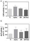
(a) LDH (lactate dehydrogenase) activity and (b) ALP (Alkaline phosphatase) activity measured in the wound fluid that was collected from the implant site after removing the implant. Values represent mean ± SEM, n = 6. No statistical differences were found between the groups.
Figure 6
Figure 6
In vivo relative mRNA expression levels of markers related to (a) inflammation: the tumor necrosis factor-α (Tnf-α) and interleukin 10 (Il-10) (b) bone resorption: tartrate-resistant acid phosphatase (Trap) and calcitonin receptor (CalcR) were analyzed as phenotypic markers, and cathepsin K (Ctsk), vacuolar type proton ATPase (H+ATPase) and metalloproteinase 9 (Mmp9) as functional osteoclastic markers and, (c) osteoblast-osteoclast interaction: osteoprotegerin (Opg) and RankL, determined by real time RT-PCR from peri-implant bone tissue on the different quercitrin implant surfaces after 8 weeks of healing. Values represent mean ± SEM, n = 6, a.u. (arbitrary units). The t-test was performed for all markers except for RankL which was analyzed by the Mann-Whitney test: * p < 0.05 vs. Ti, # p < 0.05 vs. APTES.

Similar articles

Cited by

References

    1. Moraschini V., Poubel L.A., Ferreira D.C., Barboza V.F., Barboza Edos S. Evaluation of survival and success rates of dental implants reported in longitudinal studies with a follow-up period of at least 10 years: A systematic review. Int. J. Oral Maxillofac. Surg. 2015;44:377–388. doi: 10.1016/j.ijom.2014.10.023. - DOI - PubMed
    1. Jung R.E., Pjetursson B.E., Glauser R., Zembic A., Zwahlen M., Lang N.P. A systematic review of the 5-year survival and complication rates of implant-supported single crowns. Clin. Oral Implants Res. 2008;19:119–130. doi: 10.1111/j.1600-0501.2007.01453.x. - DOI - PubMed
    1. Mombelli A., Müller N., Cionca N. The epidemiology of peri-implantitis. Clin. Oral Implants Res. 2012;23(Suppl. 6):67–76. doi: 10.1111/j.1600-0501.2012.02541.x. - DOI - PubMed
    1. Junker R., Dimakis A., Thoneick M., Jansen J.A. Effects of implant surface coatings and composition on bone integration: A systematic review. Clin. Oral Implants Res. 2009;20(Suppl. 4):185–206. doi: 10.1111/j.1600-0501.2009.01777.x. - DOI - PubMed
    1. Bauer S., Schmuki P., von der Mark K., Park J. Engineering biocompatible implant surfaces. Part I: Materials and surfaces. Prog. Mater. Sci. 2013;58:261–326. doi: 10.1016/j.pmatsci.2012.09.001. - DOI

MeSH terms

LinkOut - more resources