Review
. 2018 Dec;94(6):1073-1086.
doi: 10.1016/j.kint.2018.06.034. Epub 2018 Oct 23.

Kidney-on-a-chip: untapped opportunities

Affiliations
Review

Kidney-on-a-chip: untapped opportunities

Nureddin Ashammakhi et al. Kidney Int. 2018 Dec.

Abstract

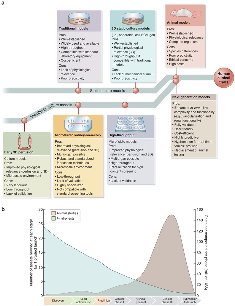
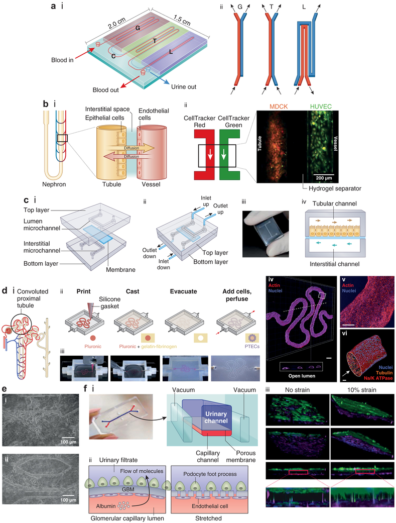
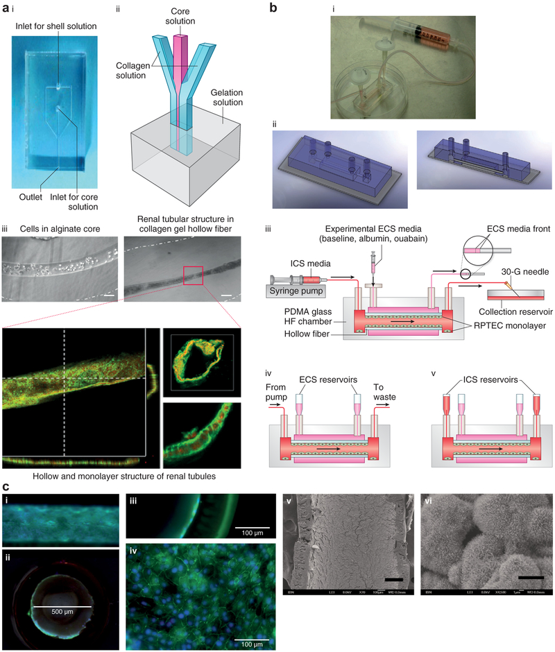
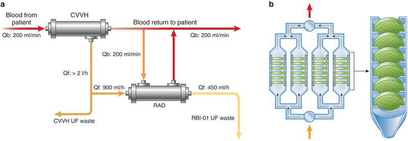
The organs-on-a-chip technology has shown strong promise in mimicking the complexity of native tissues in vitro and ex vivo, and recently significant advances have been made in applying this technology to studies of the kidney and its diseases. Individual components of the nephron, including the glomerulus, proximal tubule, and distal tubule/medullary collecting duct, have been successfully mimicked using organs-on-a-chip technology and yielding strong promises in advancing the field of ex vivo drug toxicity testing and augmenting renal replacement therapies. Although these models show promise over 2-dimensional cell systems in recapitulating important nephron features in vitro, nephron functions, such as tubular secretion, intracellular metabolism, and renin and vitamin D production, as well as prostaglandin synthesis are still poorly recapitulated in on-chip models. Moreover, construction of multiple-renal-components-on-a-chip models, in which various structures and cells of the renal system interact with each other, has remained a challenge. Overall, on-chip models show promise in advancing models of normal and pathological renal physiology, in predicting nephrotoxicity, and in advancing treatment of chronic kidney diseases.

Keywords: kidney; microfluidics; microphysiological systems; organ-on-a-chip; tissue engineering.


Figures

Figure 1 ∣
Figure 1 ∣. Future of drug testing.
(a) Evolution of 3-dimensional (3D) physiologically relevant microfluidic models for nephrotoxicity screening. ECM, extracellular matrix. Adapted with permission from Wilmer MJ, Ng CP, Lanz HL, et al. Kidney-on-a-chip technology for drug-induced nephrotoxicity screening. Trends Biotechnol. 2016;34:156–170. (b) Drug testing challenge and dilemma. Reproduced from Dehne EM, Hasenberg T, Marx U. The ascendance of microphysiological systems to solve the drug testing dilemma. Future Sci OA. 2017;3:FSO185. This work is licensed under the Creative Commons Attribution 4.0 License.
Figure 2 ∣
Figure 2 ∣. Biomimetic nephron and its components.
(a) Design of a nephron-on-a-chip model composed of an inlet for blood, an outlet for blood, and an outlet for urine. (i) Schematic showing the glomerulus (G) part, renal tubule (T) part, loop of Henle (L) part, and a connector (C) part. (ii) Schematic showing the arrangement of channels in different parts of the nephron model. Adapted from Weinberg E, Kaazempur-Mofrad M, Borenstein J. Concept and computational design for a bioartificial nephron-on-a-chip. Int J Artif Organs. 2008;31:508. (b) Biomimetic nephron (nephron-on-a-chip) using renal epithelial cells and endothelial cells (ECs) cultured in a microtubular system resembling the nephron (proximal tubule and neighboring vessel). (i) Schematic of normal structures of the nephron. Adapted from Mu X, Zheng W, Xiao L, et al. Engineering a 3D vascular network in hydrogel for mimicking a nephron. Lab Chip. 2013;13:1612, with permission of The Royal Society of Chemistry. (ii) Madin-Darby canine kidney (MDCK) cells and human renal epithelial cells (HUVECs) seeded into hydrogel microchannels as a way of mimicking the tubular and vascular proximity. Each microchannel is perfused with dye (red for MDCK and green for HUVEC). Reproduced with permission from Mu X, Zheng W, Xiao L, et al. Engineering a 3D vascular network in hydrogel for mimicking a nephron. Lab Chip. 2013;13:1612. (c) Multilayered chip, resembling renal proximal tubule (PT) composed of cells cultured on a porous membrane that separates microchannels, simulating the tubular lumen and the interstitium. The membrane is coated with either fibronectin, laminin, or Matrigel. (i) Two overlapped polydimethylsiloxane (PDMS) layers are engraved with microchannels. The apical aspect of the cells is exposed (upper channel), and the interstitium is in contact with basolateral membranes (lower channel). The channels are continuously fed in a countercurrent manner. (ii) Once assembled, the device presents 2 inlets and 2 outlet ports. (iii) Photograph of the device. (i–iii) Reproduced with permission from Sciancalepore AG, Sallustio F, Girardo S, et al. A bioartificial renal tubule device embedding human renal stem/progenitor cells. PLoS One. 2014;9:e87496. Copyright © Sciancalepore et al. (iv) Schematic illustrating the 2 channels with countercurrent flow in the 2 microchannels separated by the membrane. Adapted with permission from PLoS One. (d) (i) Schematic of the proximal convoluted tubule (PCT). (ii, iii) Fabrication of PCT steps in which a fugitive ink is printed on a gelatin-fibrinogen extracellular matrix (ECM), an additional ECM is cast around, the fugitive ink is evacuated to create a tubule, and PT cells are seeded within the tubule. (iv) A 3-dimensional (3D) rendering of the printed PCT acquired by confocal microscopy (actin is in red and nuclei in blue). A cross-sectional view is shown below, where PT cells circumscribe the open lumens in 3D. Bar = 500 μm. (v) Higher-magnification view of the region in (iv) denoted by the white rectangle. Bar = 200 μm. (vi) 3D rendering of the PCT, where an open lumen circumscribed with an epithelial lining is directionally perfused on a chip (Na/K ATPase is in red, acetylated tubulin is orange [highlighting the primary cilia], and nuclei are in blue). Bar = 50 μm. Reproduced with permission from Homan KA, Kolesky DB, Skylar-Scott MA, et al. Bioprinting of 3D convoluted renal proximal tubules on perfusable chips. Sci Rep. 2016;6:34845. Copyright © Nature Publishing Group. (e) Fibronectin-coated microchannels in PDMS chips having MDCK cells cultured inside, after 72 hours of perfusion, corresponding to 96 hours of culture (i, ii). Cells formed 3D-like tissue structures covering the microchannels. Bar = 100 μm. Reproduced with permission from Baudoin R, Griscom L, Monge M, et al. Development of a renal microchip for in vitro distal tubule models. Biotechnol Progr. 2007;23:1245–1253. Copyright © 2007 American Institute of Chemical Engineers (AIChE). (f) Images of a glomerulus-on-a-chip and schematic of the device, (i) in which terminally differentiated podocytes derived from human induced pluripotent stem cells are cultured on one side of a laminin-coated membrane and ECs on the other side in a microchannel microfluidic system, showing movement of molecules and pores appearing in the membrane upon stretching (ii). (iii) 3D reconstructed views of the tissue-tissue interface formed by podocytes (top: green) and ECs (bottom: magenta) showing that the cyclic application of 10% strain enhances the extension of podocyte cell processes through the pores of the flexible ECM-coated PDMS membrane so that they insert into the abluminal surface of the underlying EC (insets). Bar = 100 μm. Reproduced by permission from Macmillan Publishers Ltd: Nature Biomedical Enginerring, Musah S, Mammoto A, Ferrante TC, et al. Mature induced-pluripotent-stem-cell-derived human podocytes reconstitute kidney glomerular-capillary-wall function on a chip. Nat Biomed Eng. 2017;1:0069. Copyright © 2017. To optimize viewing of this image, please see the online version of this article at www.kidney-international.org.
Figure 3 ∣
Figure 3 ∣. Hollow fibers (HFs) and lab-on-a-chip.
(a) Image (i) and schematic (ii) of the collagen gel HF microfluidic device. (iii) Human kidney-2 cell-laden collagen HF with Ca-alginate core having a diameter of 100 μm (left) and HF having an inner diameter of 50 μm (right) after 10 days of culture. Confocal images of renal tubules in HF are shown at the bottom, which depicts renal tubules with a cross-sectional view (bar = 50 μm) (lower left); 3D reconstructed confocal image (bar = 50 μm) (upper right); and magnified view showing the cell-cell connection (bar = 20 μm) (lower right). Reproduced with permission from Shen C, Zhang G, Wang Q, Meng Q. Fabrication of collagen gel hollow fibers by covalent cross-linking for construction of bioengineering renal tubules. ACS Appl Mater Interf. 2015;7:19789. (b) Combining polyethersulfone-polyvinyl pyrrolidone HF and lab-on-a-chip technology. Human proximal tubular epithelial cells are cultured on the coarse inner surface of hollow fibers coated with fibrin that is embedded in a polydimethylsiloxane-glass chamber. (i) Image and (ii) schematic of the lab-on-a-chip HF bioreactor. (iii) Setup for inulin recovery perfusion studies, (iv) normal perfusion culture, and (v) static configuration for urea, creatinine, and glucose transport studies. ECS, extracapillary space surrounding the hollow fibers; ICS, intracapillary of the hollow fibers; PDMA, poly(N,N-dimethylacrylamide); RPTEC, renal proximal tubule epithelial cells. (c) Immunofluorescence and electron microscopic images of the cell-laden HF: (i) transverse section and (ii) cross-section (bar = 500 μm). Close-up views of the (iii) cross-section and (iv) transverse section (confocal image) revealing a cell monolayer in the HF (bar = 100 μm). (v) Scanning electron microscopic image of the cell-laden HF (bar = 200 μm), with (vi) the magnified view showing microvilli expression on the cell surface (bar = 5 μm). (b,c) Reproduced with permission from Ng CP, Zhuang Y, Lin AWH, Teo JCM. A fibrin-based tissue-engineered renal proximal tubule for bioartificial kidney devices: development, characterization and in vitro transport study. Int J Tissue Eng. 2013; article ID 319476. To optimize viewing of this image, please see the online version of this article at http://www.kidney-international.org.
Figure 4 ∣
Figure 4 ∣. Cell-based renal tubule assist device (RAD).
(a) Schematic of the extracorporeal perfusion circuit for cell-based RAD composed of a hemofilter perfusion pump system (continuous venovenous hemofiltration [CVVH]), a RAD perfusion system i.v. pump for the pre-RAD ultrafiltrate (UF) line, and a blood pump for the post-RAD blood line. Qb, blood flow; Qf, rate of fluid filtration. Adapted from Tumlin J. Efficacy and safety of renal tubule cell therapy for acute renal failure. J Am Soc Nephrol. 2008;19:1034–1040. (b) Bioartificial renal epithelial cell system composed of housing pieces and 4 porous disk columns, each consisting of six 2-mm-thick disks.

References

    1. Hegab HM, Elmekawy A, Stakenborg T. Review of microfluidic microbioreactor technology for high-throughput submerged microbiological cultivation. Biomicrofluidics. 2013;7:21502. - PMC - PubMed
    1. Sanechika N, Sawada K, Usui Y, et al. Development of bioartificial renal tubule devices with lifespan-extended human renal proximal tubular epithelial cells. Nephrol Dial Transplant. 2011;26:2761–2769. - PubMed
    1. Mackay S, Funke A, Buffington D, Humes H. Tissue engineering of a bioartificial renal tubule. ASAIO J. 1998;44:179–183. - PubMed
    1. Jang K, Suh K. A multi-layer microfluidic device for efficient culture and analysis of renal tubular cells. Lab Chip. 2010;10:36–42. - PubMed
    1. Hartung T. Toxicology for the twenty-first century. Nature. 2009;460:208–212. - PubMed

Publication types