. 2018 Dec 21;293(51):19797-19811.
doi: 10.1074/jbc.RA118.002742. Epub 2018 Oct 26.

FAM210B is an erythropoietin target and regulates erythroid heme synthesis by controlling mitochondrial iron import and ferrochelatase activity

Affiliations

FAM210B is an erythropoietin target and regulates erythroid heme synthesis by controlling mitochondrial iron import and ferrochelatase activity

Yvette Y Yien et al. J Biol Chem. .

Abstract

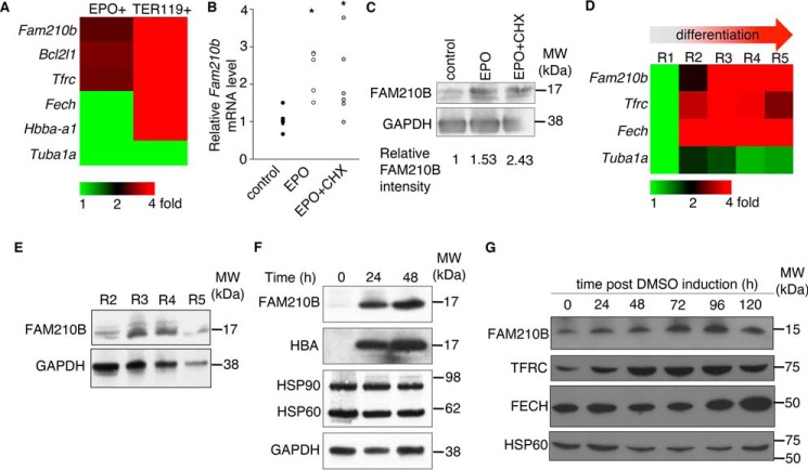
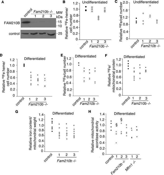
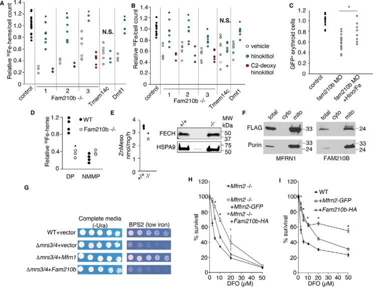
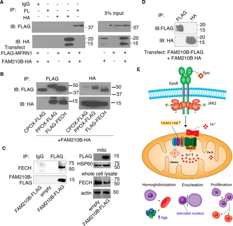
Erythropoietin (EPO) signaling is critical to many processes essential to terminal erythropoiesis. Despite the centrality of iron metabolism to erythropoiesis, the mechanisms by which EPO regulates iron status are not well-understood. To this end, here we profiled gene expression in EPO-treated 32D pro-B cells and developing fetal liver erythroid cells to identify additional iron regulatory genes. We determined that FAM210B, a mitochondrial inner-membrane protein, is essential for hemoglobinization, proliferation, and enucleation during terminal erythroid maturation. Fam210b deficiency led to defects in mitochondrial iron uptake, heme synthesis, and iron-sulfur cluster formation. These defects were corrected with a lipid-soluble, small-molecule iron transporter, hinokitiol, in Fam210b-deficient murine erythroid cells and zebrafish morphants. Genetic complementation experiments revealed that FAM210B is not a mitochondrial iron transporter but is required for adequate mitochondrial iron import to sustain heme synthesis and iron-sulfur cluster formation during erythroid differentiation. FAM210B was also required for maximal ferrochelatase activity in differentiating erythroid cells. We propose that FAM210B functions as an adaptor protein that facilitates the formation of an oligomeric mitochondrial iron transport complex, required for the increase in iron acquisition for heme synthesis during terminal erythropoiesis. Collectively, our results reveal a critical mechanism by which EPO signaling regulates terminal erythropoiesis and iron metabolism.

Keywords: cell metabolism; erythrocyte; erythropoiesis; heme; iron metabolism; red blood cell.


Conflict of interest statement

The authors declare that they have no conflicts of interest with the contents of this article

Figures

Figure 1.
Figure 1.
Fam210b is an EPO early response gene and is induced in terminally differentiating erythroid cells. A, microarray analysis of EPO-treated 32D pro-B cells shows that Fam210b is an EPO-responsive gene that is also highly enriched in the terminally differentiating TER119+ population of fetal liver erythroid cells (34). B, qRT-PCR demonstrates that Epo treatment of the EpoR-expressing pro-B cell line, 32D, up-regulated expression of Fam210b mRNA. This up-regulation persists in the presence of cycloheximide (CHX), an inhibitor of protein translation, demonstrating that Fam210b is an EPO early response gene n = 6. *, p < 0.05, Student's t test. C, FAM210B protein levels are up-regulated in response to EPO treatment of the EpoR-expressing 32D pro-B cell line. The increase in protein levels persist with cycloheximide treatment, indicating increased stability. Changes in FAM210B protein expression normalized to GAPDH are quantitated relative to control levels. D, RNAseq analysis of primary murine fetal liver cells sorted according to TER119 and CD71 (R1–R5) expression demonstrates up-regulation of Fam210b during the R2–R3 transition. E, this up-regulation is recapitulated by Western blot analysis of FAM210B protein expression. F, FAM210B protein expression is up-regulated during in vitro differentiation of primary fetal liver cells. G, FAM210B protein is induced upon terminal differentiation of MEL cells in parallel with genes required for heme synthesis, TFRC and FECH. *, p < 0.05, Student t test.
Figure 2.
Figure 2.
Expression of Fam210b is enriched in vertebrate tissues with high heme content. A, Fam210b expression (red pseudocolor) is enriched in the murine fetal liver (FL) at E12.5. B, in murine tissues, Fam210b expression is enriched in the bone marrow and fetal liver (erythroid), adult liver, skeletal muscle, and testis, all of which require high levels of iron for their function. C, zebrafish fam210b is maternally expressed during early embryonic development. At 24 hpf, fam210b expression is enriched in neural tissue and the intermediate cell mass, the site of primitive hematopoiesis (top). The expression of fam210b in the ICM is abolished in cloche embryos, which do not form hematopoietic or vascular tissue. In contrast, fam210b expression in the intermediate cell mass is expanded in dino embryos, which exhibit a ventralized phenotype (bottom), paralleling the increased expression of erythroid gata1. D, at 72 hpf, fam210b expression persists in the neural tissue and is enriched in the liver, delineated by sid4 expression. n = 3.
Figure 3.
Figure 3.
Fam210b is continuously required for terminal erythroid differentiation. A, knockdown of fam210b in zebrafish embryos with two independent antisense morpholinos caused a defect in erythroid hemoglobinization. B, Tg(globin LCR:eGFP), fam210b morphant zebrafish exhibited a decrease in GFP+ erythroid cells, indicating defective erythropoiesis, n = 4; *, p value < 0.05 Student's t test. C, shRNA-mediated knockdown of Fam210b in primary fetal liver cells decreased FAM210B protein expression. D, Fam210b knockdown primary fetal liver cells had a decrease in hemoglobin content. E, hemoglobinization defect in murine Fam210b knockdown cells is complemented by human FAM210B. F, Fam210b knockdown in primary fetal liver cells caused a cell proliferation defect. G, Fam210b knockdown caused an enucleation defect, indicative of erythroid maturation arrest. n = 3, *, p value < 0.05 Student's t test.
Figure 4.
Figure 4.
FAM210B localizes to the mitochondrial inner membrane. A, confocal fluorescence microscopy of exogenously expressed FAM210B-GFP (green) in HEK293T cells indicated that FAM210B co-localized with COXIV (red), a mitochondrial resident protein. The overlap is indicated in the merge panel (yellow). B, subcellular fractionation of primary fetal liver cells showed that FAM210B co-sedimented with HSP60 (mitochondrial) but not with HBA and GAPDH (cytoplasmic), indicating its mitochondrial localization. C, isolated mitochondria (Mito, T; lanes 1–3) purified from a MEL cell line stably expressing mouse Fam210b, was treated with proteinase K, which degraded outer membrane proteins (TOMM20) but not intermembrane space proteins (YME1L) or matrix (PreP) proteins (lane 2). Inner mitochondrial proteins, which were impervious to proteinase K treatment, (P) were separated from S proteins by centrifugation. FAM210B co-sedimented with the pellet fraction (lane 2). Mitoplasts (lanes 4–11) were generated by subjecting intact mitochondria to osmotic shock, exposing inner membrane and intermembrane space proteins to proteinase K digestion. Mitoplasts were centrifuged to separate the soluble proteins from the mitoplast pellet. Proteinase K treatment degraded a fraction of FAM210B (lane 6) and completely degraded YME1L but not PreP, suggesting that at least a fraction of FAM210B was situated in the mitochondrial inner membrane or intermembrane space. Triton X-100 (TX-100) treatment liberated FAM210B, PreP, and YME1L from the pellet into the soluble fraction (lane 9). These solubilized proteins were digested by proteinase K, demonstrating specificity of assay (lane 10). The asterisk marks a nonspecific band that was detected with the mouse FAM210B antibody; the triangle marks a likely FAM210B degradation product; and the square marks a core of PreP that is tightly folded and resistant to protease treatment. D, to determine whether FAM210B was an integral inner-membrane protein, isolated mitochondria (T) were analyzed by carbonate extraction. P and S fractions were analyzed by immunoblotting for FAM210B. The blot was probed for TIMM23 as an integral membrane protein control and mortalin as a soluble protein control. An asterisk marks nonspecific bands detected by the FAM210B antibody.
Figure 5.
Figure 5.
Fam210b is required for heme synthesis by facilitating mitochondrial iron import. A, Fam210b−/− MEL cell lines generated by CRISPR/Cas9 expressed no detectable FAM210B protein. B, 55Fe metabolic labeling showed that undifferentiated Fam210b−/− MEL cell clones did not cause changes in heme synthesis (n = 3) or iron uptake (n = 3) (C). D, differentiated Fam210b knockout MEL cells exhibited defective heme synthesis (n = 6). E, iron uptake (n = 5). F, mitochondria from differentiated Fam210b knockout cells had decreased 55Fe-transferrin uptake (n = 5). G, ICP-MS demonstrated that mitochondria from Fam210b knockout cells had a decrease in total iron (n = 6). H, mitochondrial aconitase activity, which depends on [Fe-S] cluster synthesis, is decreased in Fam210b−/− cells indicating a defect in mitochondrial iron acquisition. Mfrn1−/− MEL clones served as controls for mitochondrial iron deficiency (n = 4). *, p < 0.05; §, p < 0.1 Student's t test. All data are normalized to WT controls.
Figure 6.
Figure 6.
Fam210B increases the kinetics of mitochondrial iron import in differentiating MEL cells by functioning as an auxiliary factor. A, 55Fe metabolic labeling confirmed a decrease in heme synthesis in differentiating Fam210b (CRISPR1–3), Tmem14c, and Dmt1 deficient MEL cells (black bars). The addition of hinokitiol, a lipid-soluble iron carrier (white bars), restored heme synthesis in Fam210b- and Dmt1-deficient cells, but not Tmem14c-deficient cells. C2-deoxyhinokitiol, which does not chelate iron, did not complement heme synthesis in the Fam210b knockout cells (gray bars) (n = 6). B, 55Fe metabolic labeling confirmed an iron uptake defect in differentiating Fam210b-, Tmem14c-, and Dmt1-deficient cells (black bars). The iron uptake defect in Fam210b- and Dmt1-deficient cells was chemically complemented by hinokitiol (white bars). Hinokitiol did not rescue iron uptake in Tmem14c-deficient cells, which have a porphyrin synthesis defect. C2-deoxyhinokitiol did not rescue the iron deficiency in Fam210b−/− cells (gray bars) (n = 6). C, treatment of fam210b morphant zebrafish embryos (MO) with iron citrate and hinokitiol (MO + Hino/Fe) rescued their anemia (n = 8). D, FECH activity was measured in intact mitochondria isolated from WT and Fam210b−/− MEL cells, using 55Fe and DP as substrates. FECH activity in Fam210b−/− mitochondria was ∼1/3 that of WT controls. Both WT and Fam210b−/− mitochondria had very little measurable FECH activity when NMMP was used as a substrate (n = 4). E, FECH activity was measured in isolated mitochondria from WT and Fam210b−/− MEL cells that were treated with detergent and disrupted by sonication, allowing access of reaction substrates to FECH in the absence of intact membranes. FECH activity was decreased in Fam210b−/− mitochondria, but not to the extent of intact mitochondria (n = 3). F, FLAG-tagged MFRN1 and FAM210B co-localized with porin in Δmrs3/4 yeast, indicating correct mitochondrial localization. G, Mfrn1 expression complemented the growth defect of Δmrs3/4 yeast in low-iron media, whereas Fam210b expression did not. H, survival of Mfrn2−/− fibroblasts in DFO is significantly complemented by expression of Mfrn2-GFP, but less so by expression of Fam210b-HA (n = 3). I, expression of Fam210b-HA and Mfrn2-GFP in WT fibroblasts increase cell survival in the presence of DFO, with Fam210b overexpression possessing a protective effect over a larger dose range than Mfrn2-GFP. Mean ± S.E., n = 3, *, p < 0.05; §, p < 0.1 Student's t test. N.S., not significant.
Figure 7.
Figure 7.
FAM210B directly interacts with terminal heme synthesis enzymes but does not with MFRN1 (Slc25a37). A, immunoprecipitation of co-expressed FAM210B-HA and FLAG-MFRN1 demonstrates FAM210B does not directly interact with MFRN1. B, FAM210B-HA interacts with the terminal enzymes of the heme synthesis pathway, PPOX and FECH, but not CPOX. MFRN1, CPOX, PPOX, and FECH are all localized to the mitochondrial inner membrane. C, immunoprecipitation of FAM-210B FLAG stably expressed in differentiating MEL cells shows that ectopically expressed FAM210B interacts with endogenous FECH. D, FAM210B-HA and FAM210B-FLAG do not interact, demonstrating that FAM210B does not homooligomerize. E, model of the role of FAM210B as an adaptor protein in an oligomeric complex with terminal heme enzymes, required for terminal erythropoiesis and mitochondrial iron importation.

References

    1. Richmond T. D., Chohan M., and Barber D. L. (2005) Turning cells red: signal transduction mediated by erythropoietin. Trends Cell Biol. 15, 146–155 10.1016/j.tcb.2005.01.007 - DOI - PubMed
    1. Gammella E., Diaz V., Recalcati S., Buratti P., Samaja M., Dey S., Noguchi C. T., Gassmann M., and Cairo G. (2015) Erythropoietin's inhibiting impact on hepcidin expression occurs indirectly. Am. J. Physiol. Regul. Integr. Comp. Physiol. 308, R330–R335 10.1152/ajpregu.00410.2014 - DOI - PMC - PubMed
    1. Nai A., Rubio A., Campanella A., Gourbeyre O., Artuso I., Bordini J., Gineste A., Latour C., Besson-Fournier C., Lin H. Y., Coppin H., Roth M.-P., Camaschella C., Silvestri L., Meynard D., Franke K., et al. (2016) Limiting hepatic Bmp-Smad signaling by matriptase-2 is required for erythropoietin-mediated hepcidin suppression in mice. Blood 127, 99–101 10.1182/blood-2015-11-681494 - DOI - PMC - PubMed
    1. Feng D., and Lazar M. A. (2012) Clocks, metabolism, and the epigenome. Mol. Cell 47, 158–167 10.1016/j.molcel.2012.06.026 - DOI - PMC - PubMed
    1. Girvan H. M., and Munro A. W. (2013) Heme sensor proteins. J. Biol. Chem. 288, 13194–13203 10.1074/jbc.R112.422642 - DOI - PMC - PubMed

Publication types