. 2018 Dec 13;85(1):e01938-18.
doi: 10.1128/AEM.01938-18. Print 2019 Jan 1.

Siderophore-Mediated Iron Acquisition Enhances Resistance to Oxidative and Aromatic Compound Stress in Cupriavidus necator JMP134

Affiliations

Siderophore-Mediated Iron Acquisition Enhances Resistance to Oxidative and Aromatic Compound Stress in Cupriavidus necator JMP134

Changfu Li et al. Appl Environ Microbiol. .

Abstract

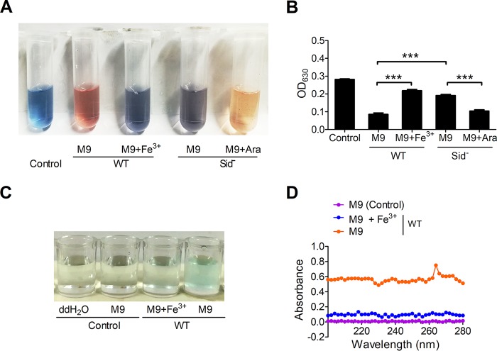
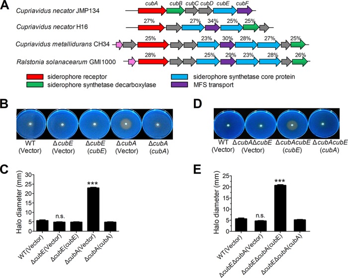
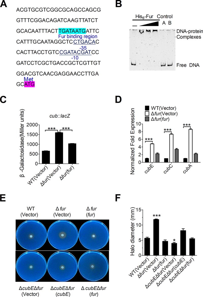
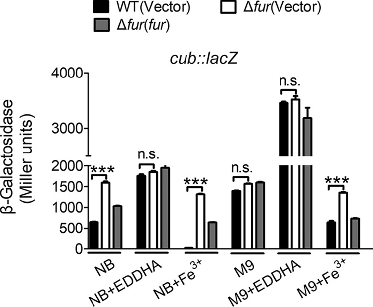
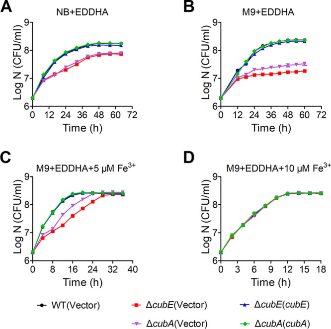
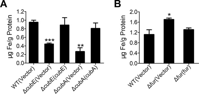
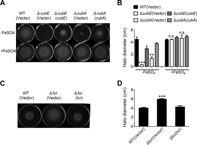
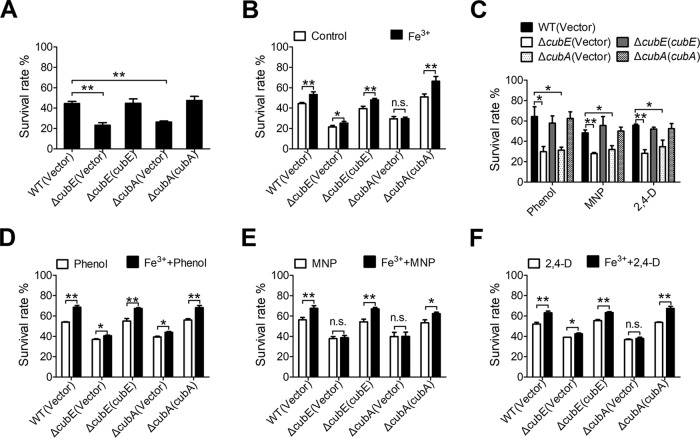
Many bacteria secrete siderophores to enhance iron uptake under iron-restricted conditions. In this study, we found that Cupriavidus necator JMP134, a well-known aromatic pollutant-degrading bacterium, produces an unknown carboxylate-type siderophore named cupriabactin to overcome iron limitation. Using genome mining, targeted mutagenesis, and biochemical analysis, we discovered an operon containing six open reading frames (cubA-F) in the C. necator JMP134 genome that encodes proteins required for the biosynthesis and uptake of cupriabactin. As the dominant siderophore of C. necator JMP134, cupriabactin promotes the growth of C. necator JMP134 under iron-limited conditions via enhanced ferric iron uptake. Furthermore, we demonstrated that the iron concentration-dependent expression of the cub operon is mediated by the ferric uptake regulator (Fur). Physiological analyses revealed that the cupriabactin-mediated iron acquisition system influences swimming motility, biofilm formation, and resistance to oxidative and aromatic compound stress in C. necator JMP134. In conclusion, we identified a carboxylate-type siderophore named cupriabactin, which plays important roles in iron scavenging, bacterial motility, biofilm formation, and stress resistance.IMPORTANCE Since siderophores have been widely exploited for agricultural, environmental, and medical applications, the identification and characterization of new siderophores from different habitats and organisms will have great beneficial applications. Here, we identified a novel siderophore-producing gene cluster in C. necator JMP134. This gene cluster produces a previously unknown carboxylate siderophore, cupriabactin. Physiological analyses revealed that the cupriabactin-mediated iron acquisition system influences swimming motility, biofilm formation, and oxidative stress resistance. Most notably, this system also plays important roles in increasing the resistance of C. necator JMP134 to stress caused by aromatic compounds, which provide a promising strategy to engineer more efficient approaches to degrade aromatic pollutants.

Keywords: Cupriavidus necator; Fur; aromatic compound degradation; biofilm; oxidative stress; siderophore.


Figures

FIG 1
FIG 1
Presence of a carboxylate siderophore in C. necator JMP134. (A and B) Detection of siderophore production in C. necator JMP134 wild type (WT) and the Sid mutant cultured in M9 medium with or without Fe3+ (100 μM) using the CAS assay. (A) The CAS solution takes on blue, as CAS, HTDMA (hexadecyltrimethylammonium bromide), and Fe(III) can form a triple FeDye3-x complex which has an adsorption at 630 nm. When siderophore removes Fe(III) from the complex, the color of the solution will turn to red or brown. Arabinose (Ara; 0.2%) was supplemented to the medium to induce the expression of the siderophore biosynthesis genes. (B) Quantification of blue FeDye3-x complex formation in panel A by determining the absorbance at 630 nm. The decreasing of absorbance indicates the production of siderophore. (C and D) Detection of carboxylate siderophore in C. necator JMP134 wild type cultured in M9 medium with or without Fe3+ (100 μM) by using the copper sulfate reagent. Siderophore can form a blue complex (C) with copper sulfate, and the absorbance was recorded at 264 nm (D). Data represent the mean ± SEM of three biological replicates, each of which was performed in three technical replicates. ***, P < 0.001. ddH2O, double-distilled water.
FIG 2
FIG 2
Siderophore production from the cub operon. (A) Comparison of the cupriabactin biosynthesis genes from C. necator JMP134 with the staphyloferrin B biosynthesis-related genes from indicated strains. Numbers above each ORF indicate the amino acid identity (expressed as a percentage) to the corresponding ORF of C. necator JMP134. The GenBank accession numbers are as follows: Cupriavidus pinatubonensis JMP134, AAZ63039.1 to AAZ63041.1; Cupriavidus necator H16, AAP85873.1 to AAP85879.1; Cupriavidus metallidurans CH34, ABF07996.1 to ABF08006.1; and Ralstonia solanacearum GMI1000, CAD17566.1 to CAD17575.1. (B and C) Siderophore-mediated iron acquisition requires cubE and cubA. (B) The production of siderophore was detected using colorimetric CAS agar plates. For each strain, stationary-phase strains were taken from a single colony grown on nutrient agar, spotted onto the middle of the CAS plates, and photographed after incubation for 5 days at 30°C. (C) The diameter of the halo of color change was measured at its widest point from the edge of colony growth. The graph represents the mean halo diameter from three individual plates for each strain. (D and E) Comparison of the colored halos around C. necator JMP134 wild type, ΔcubE ΔcubA double mutant, and ΔcubE ΔcubA double mutant strains complemented with cubE or cubA, respectively. (E) The graph represents the mean halo diameter from three individual plates for each strain. The vector stands for the pBBR1MCS-2 plasmid. Data represent the mean ± SEM of three biological replicates, each of which was performed with three technical replicates. ***, P < 0.001; n.s., not significant.
FIG 3
FIG 3
Fur negatively regulates cub expression. (A) Identification of a Fur-binding site in the promoter region of cub. The putative Fur-binding site identified by the online software Virtual Footprint is indicated by blue highlighting. The ATG start codon of the first ORF of the cub operon is shown. Putative −35 and −10 elements of the cub promoter are underlined. (B) EMSA was performed to analyze the interactions between His6-Fur and the promoter. Increasing amounts of Fur (0.38, 0.76, and 1.14 μM) and 0 to 1 nM the DNA fragment were used. As negative controls, a 75-bp unrelated DNA fragment (control A) and 1.14 μM BSA (control B) were included in this assay. (C) Fur represses the expression of cub. β-Galactosidase analyses of cub promoter activities were performed using the transcriptional Pcub::lacZ chromosomal fusion reporter expressed in the C. necator JMP134 wild-type, Δfur mutant, and complemented Δfur(fur) strains grown to stationary phase in NB medium. (D) qRT-PCR analysis of mRNA levels of cub. Cells of relevant C. necator JMP134 strains were grown to mid-exponential phase in NB medium, and the expression of cubE, cubC, and cubA (the main components of cub) were measured by qRT-PCR. (E and F) Siderophore production is negatively regulated by Fur. (E) The production of siderophore was detected using colorimetric CAS agar plates to compare the colored halos around wild-type, Δfur mutant, the complemented Δfur(fur) mutant, ΔcubE Δfur double mutants, and the complemented ΔcubE Δfur(cubE) and ΔcubE Δfur(fur) mutants. (F) The graph represents the mean halo diameter from three individual plates for each strain after incubation for 5 days at 30°C. Vector stands for pBBR1MCS-5 (C) and pBBR1MCS-2 (D to F). Data represent the mean ± SEM of three biological replicates, each of which was performed in three technical replicates. ***, P < 0.001; *, P < 0.05.
FIG 4
FIG 4
Fur-mediated expression of cub responds to different iron concentrations. Relevant C. necator JMP134 wild-type, Δfur mutant, and the complemented Δfur(fur) mutant harboring Pcub::lacZ were grown in NB medium with 2.0 μM EDDHA and 20 μM Fe3+ and in M9 medium with 2.0 μM EDDHA and 20 μM Fe3+, and the expression of the reporter in each strain was measured. The vector stands for the pBBR1MCS-5 plasmid. Data represent the mean ± SEM of three biological replicates, each of which was performed in three technical replicates. ***, P < 0.001; n.s., not significant.
FIG 5
FIG 5
Growth curves of relevant C. necator JMP134 strains in NB and iron-limiting media at 30°C. (A to D) Relevant C. necator JMP134 wild-type, ΔcubE mutant, the complemented ΔcubE(cubE) mutant, ΔcubA mutant, and the complemented ΔcubA(cubA) mutant strains grown overnight in 3 ml of NB medium were harvested, washed, resuspended in M9 medium, and diluted 1:500 in fresh NB medium with 5 μM EDDHA (A), M9 medium with 5 μM EDDHA (B), M9 medium with 5 μM EDDHA and 5 μM Fe3+ (C), and M9 medium with 5 μM EDDHA and 10 μM Fe3+ (D). The growth of the cultures was monitored by measuring the OD600 at the indicated time points. IPTG (1 mM) was included in the medium for induction. Vector stands for the pBBR1MCS-2 plasmid. Data represent the mean ± SEM of three biological replicates, each of which was performed in three technical replicates.
FIG 6
FIG 6
Cupriabactin is involved in iron acquisition. (A) Iron uptake requires cupriabactin. Relevant C. necator JMP134 wild-type, ΔcubE mutant, the complemented ΔcubE(cubE) mutant, ΔcubA mutant, and the complemented ΔcubA(cubA) mutant were grown overnight in M9 medium, which was incubated to the end of logarithmic phase, harvested, and washed, and the intracellular iron associated with bacterial cells was measured by atomic absorption spectroscopy. (B) The Δfur mutant accumulates intracellular iron. Intracellular iron was measured in the C. necator JMP134 wild-type, Δfur mutant, and complemented Δfur(fur) mutant strains grown to the end of logarithmic phase in NB medium. The vector stands for the pBBR1MCS-2 plasmid. Data represent the mean ± SEM of three biological replicates, each of which was performed in three technical replicates. ***, P < 0.001; **, P < 0.01; *, P < 0.05.
FIG 7
FIG 7
Swimming motility is influenced by intracellular iron. (A and B). Iron supplementation restores motility in the ΔcubE and ΔcubA mutants. A single colony of indicated C. necator JMP134 strains was touched slightly on swimming medium with or without 20 μM FeSO4. Photographs were taken (A), and halo diameters around the colonies were measured after 32 h of incubation at 30°C (B). Experiments were performed with at least three biological replicates in five technical replicates each. (C and D) Fur regulation influences surface motility. Motility of the C. necator wild-type, Δfur mutant, the complemented Δfur(fur) mutant strains is shown on swimming plates. Photographs were taken (C), and halo diameters around the colonies were measured after 16 h of incubation at 30°C (D). Experiments were conducted in at least three biological replicates in five technical replicates each. The vector stands for the pBBR1MCS-2 plasmid. Data represent the mean ± SEM of three biological replicates, each of which was performed in five technical replicates. ***, P < 0.001; **, P < 0.01; n.s., not significant.
FIG 8
FIG 8
Effect of cupriabactin on biofilm formation in iron-limiting media. (A and B) Overnight bacterial cultures were diluted 100-fold in fresh NB medium containing 1 mM IPTG and 50 µg ml−1 kanamycin, in the absence and presence of 2.0 μM EDDHA. After vertical incubation for 5 days with shaking at 140 rpm in 30°C, biofilm formation of the strains were determined by crystal violet staining (A) and quantified using optical density measurement (B). The vector stands for the pBBR1MCS-2 plasmid. Data represent the mean ± SEM of three biological replicates, each in three technical replicates. ***, P < 0.001; n.s., not significant.
FIG 9
FIG 9
Cupriabactin-mediated active iron acquisition contributes to stress resistance. (A) Alleviation of the sensitivity of C. necator JMP134 strains to H2O2 required the CubE and CubA proteins. The viability of mid-exponential-phase C. necator JMP134 strains was determined after exposure to H2O2 for 25 min. (B) Alleviation of the sensitivity of C. necator JMP134 strains to H2O2 by exogenous Fe3+ (1.5 μM). Indicated bacterial strains grown to mid-exponential phase were exposed to H2O2 with 1.5 μM Fe3+ for 25 min, and the viability of the cells was determined. (C) Alleviation of the sensitivity of C. necator JMP134 strains to phenol, MNP, and 2,4-D required the CubE and CubA proteins. Indicated bacterial strains grown to mid-exponential phase were exposed to phenol (30 mM), MNP (3 mM), or 2,4-D (20 mM) for 25 min, and the viability of the cells was determined. (D to F) Alleviation of the sensitivity of C. necator JMP134 strains to phenol, MNP and 2,4-D by exogenous Fe3+ (1.5 μM). Indicated bacterial strains grown to mid-exponential phase were exposed to phenol (D), MNP (E), or 2,4-D (F) with 1.5 μM Fe3+ for 25 min, and the viability of the cells was determined. The vector stands for the pBBR1MCS-2 plasmid. Data represent the mean ± SEM of three biological replicates, each of which was performed in three technical replicates. **, P < 0.01; *, P < 0.05; n.s., not significant.

Similar articles

Cited by

References

    1. Miethke M, Marahiel MA. 2007. Siderophore-based iron acquisition and pathogen control. Microbiol Mol Biol Rev 71:413–451. doi:10.1128/MMBR.00012-07. - DOI - PMC - PubMed
    1. Tanabe T, Naka A, Aso H, Nakao H, Narimatsu S, Inoue Y, Ono T, Yamamoto S. 2005. A novel aerobactin utilization cluster in Vibrio vulnificus with a gene involved in the transcription regulation of the iutA homologue. Microbiol Immunol 49:823–834. doi:10.1111/j.1348-0421.2005.tb03671.x. - DOI - PubMed
    1. Hider RC, Kong X. 2010. Chemistry and biology of siderophores. Nat Prod Rep 27:637–657. doi:10.1039/b906679a. - DOI - PubMed
    1. Kurth C, Kage H, Nett M. 2016. Siderophores as molecular tools in medical and environmental applications. Org Biomol Chem 14:8212–8227. doi:10.1039/c6ob01400c. - DOI - PubMed
    1. Schaible UE, Kaufmann SH. 2004. Iron and microbial infection. Nat Rev Microbiol 2:946–953. doi:10.1038/nrmicro1046. - DOI - PubMed

Publication types

LinkOut - more resources