

Genomic Characterization of Cyanophage vB_AphaS-CL131 Infecting Filamentous Diazotrophic Cyanobacterium Aphanizomenon flos-aquae Reveals Novel Insights into Virus-Bacterium Interactions
- PMID: 30367000
- PMCID: PMC6293099
- DOI: 10.1128/AEM.01311-18
Genomic Characterization of Cyanophage vB_AphaS-CL131 Infecting Filamentous Diazotrophic Cyanobacterium Aphanizomenon flos-aquae Reveals Novel Insights into Virus-Bacterium Interactions
Abstract
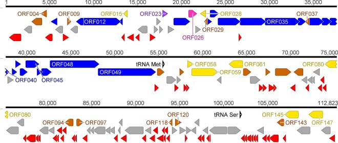
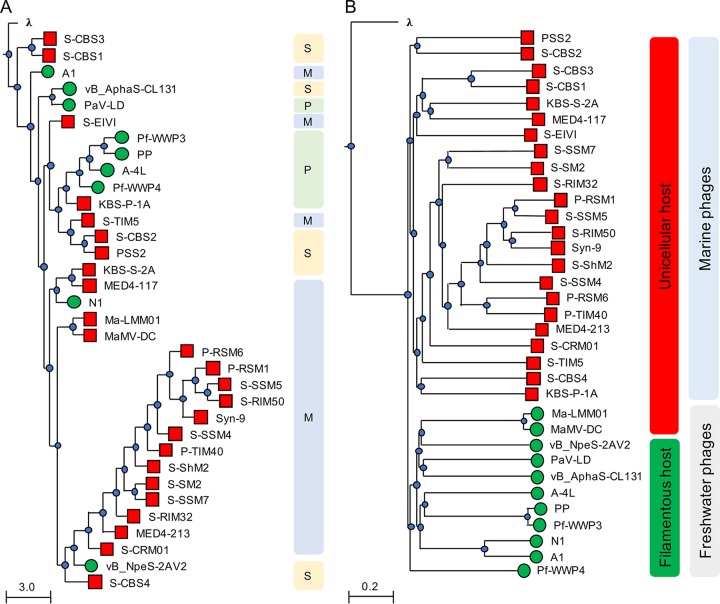
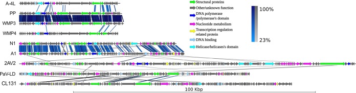
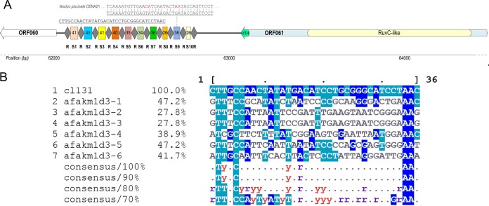
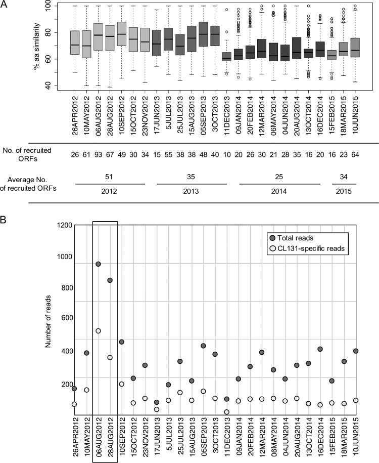
While filamentous cyanobacteria play a crucial role in food web dynamics and biogeochemical cycling of many aquatic ecosystems around the globe, the knowledge regarding the phages infecting them is limited. Here, we describe the complete genome of the virulent cyanophage vB_AphaS-CL131 (here, CL 131), a Siphoviridae phage that infects the filamentous diazotrophic bloom-forming cyanobacterium Aphanizomenon flos-aquae in the brackish Baltic Sea. CL 131 features a 112,793-bp double-stranded DNA (dsDNA) genome encompassing 149 putative open reading frames (ORFs), of which the majority (86%) lack sequence homology to genes with known functions in other bacteriophages or bacteria. Phylogenetic analysis revealed that CL 131 possibly represents a new evolutionary lineage within the group of cyanophages infecting filamentous cyanobacteria, which form a separate cluster from phages infecting unicellular cyanobacteria. CL 131 encodes a putative type V-U2 CRISPR-Cas system with one spacer (out of 10) targeting a DNA primase pseudogene in a cyanobacterium and a putative type II toxin-antitoxin system, consisting of a GNAT family N-acetyltransferase and a protein of unknown function containing the PRK09726 domain (characteristic of HipB antitoxins). Comparison of CL 131 proteins to reads from Baltic Sea and other available fresh- and brackish-water metagenomes and analysis of CRISPR-Cas arrays in publicly available A. flos-aquae genomes demonstrated that phages similar to CL 131 are present and dynamic in the Baltic Sea and share a common history with their hosts dating back at least several decades. In addition, different CRISPR-Cas systems within individual A. flos-aquae genomes targeted several sequences in the CL 131 genome, including genes related to virion structure and morphogenesis. Altogether, these findings revealed new genomic information for exploring viral diversity and provide a model system for investigation of virus-host interactions in filamentous cyanobacteria.IMPORTANCE The genomic characterization of novel cyanophage vB_AphaS-CL131 and the analysis of its genomic features in the context of other viruses, metagenomic data, and host CRISPR-Cas systems contribute toward a better understanding of aquatic viral diversity and distribution in general and of brackish-water cyanophages infecting filamentous diazotrophic cyanobacteria in the Baltic Sea in particular. The results of this study revealed previously undescribed features of cyanophage genomes (e.g., self-excising intein-containing putative dCTP deaminase and putative cyanophage-encoded CRISPR-Cas and toxin-antitoxin systems) and can therefore be used to predict potential interactions between bloom-forming cyanobacteria and their cyanophages.
Keywords: Baltic Sea; Siphoviridae; TA system; brackish environment; phage-encoded CRISPR-Cas.
Copyright © 2018 Šulčius et al.
Figures
Similar articles
-
A Review of Cyanophage-Host Relationships: Highlighting Cyanophages as a Potential Cyanobacteria Control Strategy.Toxins (Basel). 2022 May 31;14(6):385. doi: 10.3390/toxins14060385. Toxins (Basel). 2022. PMID: 35737046 Free PMC article. Review.
-
Characterization of a lytic cyanophage that infects the bloom-forming cyanobacterium Aphanizomenon flos-aquae.FEMS Microbiol Ecol. 2015 Feb;91(2):1-7. doi: 10.1093/femsec/fiu012. Epub 2014 Dec 5. FEMS Microbiol Ecol. 2015. PMID: 25764544
-
Viruses Infecting a Freshwater Filamentous Cyanobacterium (Nostoc sp.) Encode a Functional CRISPR Array and a Proteobacterial DNA Polymerase B.mBio. 2016 Jun 14;7(3):e00667-16. doi: 10.1128/mBio.00667-16. mBio. 2016. PMID: 27302758 Free PMC article.
-
Nitrogen Flow in Diazotrophic Cyanobacterium Aphanizomenon flos-aquae Is Altered by Cyanophage Infection.Front Microbiol. 2020 Aug 19;11:2010. doi: 10.3389/fmicb.2020.02010. eCollection 2020. Front Microbiol. 2020. PMID: 32973727 Free PMC article.
-
A review of the phylogeny, ecology and toxin production of bloom-forming Aphanizomenon spp. and related species within the Nostocales (cyanobacteria).Harmful Algae. 2016 Apr;54:21-43. doi: 10.1016/j.hal.2015.09.007. Harmful Algae. 2016. PMID: 28073477 Review.
Cited by
-
A Novel Freshwater Cyanophage Mae-Yong1326-1 Infecting Bloom-Forming Cyanobacterium Microcystis aeruginosa.Viruses. 2022 Sep 16;14(9):2051. doi: 10.3390/v14092051. Viruses. 2022. PMID: 36146857 Free PMC article.
-
Genomic Analysis and Taxonomic Characterization of Seven Bacteriophage Genomes Metagenomic-Assembled from the Dishui Lake.Viruses. 2023 Sep 30;15(10):2038. doi: 10.3390/v15102038. Viruses. 2023. PMID: 37896815 Free PMC article.
-
Host Cyanobacteria Killing by Novel Lytic Cyanophage YongM: A Protein Profiling Analysis.Microorganisms. 2022 Jan 24;10(2):257. doi: 10.3390/microorganisms10020257. Microorganisms. 2022. PMID: 35208712 Free PMC article.
-
Novel Freshwater Cyanophages Provide New Insights into Evolutionary Relationships between Freshwater and Marine Cyanophages.Microbiol Spectr. 2021 Oct 31;9(2):e0059321. doi: 10.1128/Spectrum.00593-21. Epub 2021 Sep 29. Microbiol Spectr. 2021. PMID: 34585945 Free PMC article.
-
A Review of Cyanophage-Host Relationships: Highlighting Cyanophages as a Potential Cyanobacteria Control Strategy.Toxins (Basel). 2022 May 31;14(6):385. doi: 10.3390/toxins14060385. Toxins (Basel). 2022. PMID: 35737046 Free PMC article. Review.
References
-
- Mühling M, Fuller NJ, Millard A, Somerfield PJ, Marie D, Wilson WH, Scanlan DJ, Post AF, Joint I, Mann NH. 2005. Genetic diversity of marine Synechococcus and co-occurring cyanophage communities: evidence for viral control of phytoplankton. Environ Microbiol 7:499–508. doi:10.1111/j.1462-2920.2005.00713.x. - DOI - PubMed
-
- Weitz JS, Stock CA, Wilhelm SW, Bourouiba L, Coleman ML, Buchan A, Follows MJ, Fuhrman JA, Jover LF, Lennon JT, Middelboe M, Sonderegger DL, Suttle CA, Taylor BP, Thingstad TF, Wilson WH, Wommack KE. 2015. A multitrophic model to quantify the effects of marine viruses on microbial food webs and ecosystem processes. ISME J 9:1352–1364. doi:10.1038/ismej.2014.220. - DOI - PMC - PubMed
-
- Guidi L, Chaffron S, Bittner L, Eveillard D, Larhlimi A, Roux S, Darzi Y, Audic S, Berline L, Brum J, Coelho LP, Espinoza JCI, Malviya S, Sunagawa S, Dimier C, Kandels-Lewis S, Picheral M, Poulain J, Searson S, Coordinators TO, Stemmann L, Not F, Hingamp P, Speich S, Follows M, Karp-Boss L, Boss E, Ogata H, Pesant S, Weissenbach J, Wincker P, Acinas SG, Bork P, de Vargas C, Iudicone D, Sullivan MB, Raes J, Karsenti E, Bowler C, Gorsky G. 2016. Plankton networks driving carbon export in the oligotrophic ocean. Nature 532:465–470. doi:10.1038/nature16942. - DOI - PMC - PubMed
Publication types
MeSH terms
Substances
LinkOut - more resources
Full Text Sources
